
Toric IOL misalignment occurs when a toric intraocular lens lies away from its intended axis, and the result is residual astigmatism that undermines the purpose of the implant.
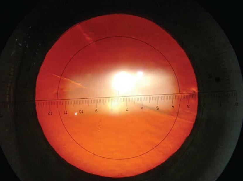
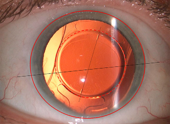
A few degrees of rotation are common and tolerable, but larger errors reduce the correction and can add astigmatism.
Recognizing the problem early gives the surgeon the chance to reposition the lens before the capsule seals it in place.
How Much Toric IOL Misalignment Matters
The effective power of the toric lens falls by about 3 percent for each degree of misalignment.
A 10-degree error leaves roughly a third of the astigmatism uncorrected, and a 30-degree error removes the benefit.
Beyond about 30 degrees the lens starts to add astigmatism, and the patient can end up with more cylinder than before surgery.
These figures are approximate, and they depend on the power of the lens, but the relationship is steep enough that axis accuracy deserves attention.
Causes
- Marking errors: incorrect reference marks on the cornea, especially with cyclotorsion when the patient lies down
- Biometry and keratometry errors: an inaccurate axis or an incorrect assessment of the posterior cornea
- Intraoperative misplacement: an error in aligning the lens in the capsular bag
- Postoperative rotation: movement of the lens in the early postoperative days, before capsular fibrosis fixes it
- Large capsular bag: an axial myopic eye with a large bag allows more movement
- Lens design and material: haptic configuration and surface properties influence stability
Most rotation takes place in the first days to weeks, and it usually stops when the capsule contracts around the lens.
All-fit Slit-Lamp Adapter
Record and share exactly what you see at the slit lamp. One adapter fits any slit lamp or surgical microscope — and any smartphone.
From Choroida — the team behind this siteDetecting Misalignment
The main symptom is blur and a refractive result that does not match the plan.
Check the uncorrected acuity, the manifest refraction, and the keratometry.
If the residual cylinder is higher than expected, dilate the pupil and examine the lens at the slit lamp with a narrow beam, noting the axis marks on the optic.
Compare the lens axis with the planned axis, using a protractor eyepiece or an image captured with a smartphone or a camera.
Online calculators can estimate the best repositioning angle based on the refraction and the current lens axis.
Take care to examine the cornea, because corneal irregularity, dry eye, or an error in the keratometry may explain the findings and would not be corrected by rotating the lens.
Management
Reposition the lens when the residual astigmatism is significant and the lens is out of axis by a meaningful amount.
The procedure is performed in the operating room, with a viscoelastic and a spatula or a hook, and it is best done within the first few weeks, before capsular fibrosis.
Later repositioning is possible, but it may require more manipulation to break the adhesions between the capsule and the lens.
If the lens is correctly placed and the astigmatism persists, consider limbal relaxing incisions or laser refractive surgery, as appropriate.
A lens exchange is reserved for cases in which the power or the position cannot be corrected.
Before surgery, patients should be told that toric implants can rotate and that a second operation is occasionally needed.
Review all measurements after surgery to improve the planning of future cases (see phacoemulsification).
Preventing Misalignment
Precise preoperative measurements are the foundation of a good toric result.
Use two independent keratometry methods and compare them, and consider the effect of the posterior corneal surface, which matters most in eyes with against-the-rule astigmatism.
Mark the cornea with the patient sitting upright to avoid errors from cyclotorsion, or use an image-guided system that registers the iris and limbal landmarks.
During the operation, remove all viscoelastic from behind the lens, because retained material allows the lens to rotate in the first postoperative hours.
Choose a lens with a stable haptic design, and take care to center it within a capsulorhexis that overlaps the optic edge.
Postoperative Checks
Examine the lens axis at the first visit and again at one to two weeks, with the pupil dilated if necessary.
If the lens has rotated and the surgeon plans to reposition it, it is easier to do so early, which is why the first examinations matter.
Patients should be told to avoid rubbing the eye and to report a sudden blur after the first weeks.
Counselling After a Disappointing Result
Explain that the astigmatism is not permanent, and that options exist for correction.
A clear plan, including the proposed surgery and the likely outcome, helps patients to remain confident in their surgeon.


Document what you see
Two smartphone imaging tools built for everyday clinic use — one for the slit lamp, one for the fundus.
From Choroida — the team behind this siteReferences
- Shimizu K, Misawa A, Suzuki Y. Toric intraocular lenses: correcting astigmatism while controlling axis. J Cataract Refract Surg. 1994;20:523-526.
- Kessel L, Andresen J, Tendal B, et al. Toric intraocular lenses in the correction of astigmatism during cataract surgery: a systematic review and meta-analysis. Ophthalmology. 2016;123:275-286.
- Salmon JF. Kanski’s Clinical Ophthalmology: A Systematic Approach. 9th ed. Elsevier; 2020.
- American Academy of Ophthalmology. Basic and Clinical Science Course, Section 11: Lens and Cataract. AAO; current edition.

Writes and reviews the clinical articles on Educate. Every post is checked against peer-reviewed sources before it goes live.


