
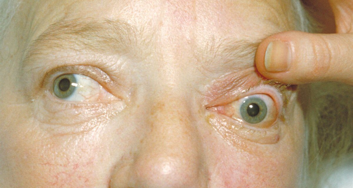
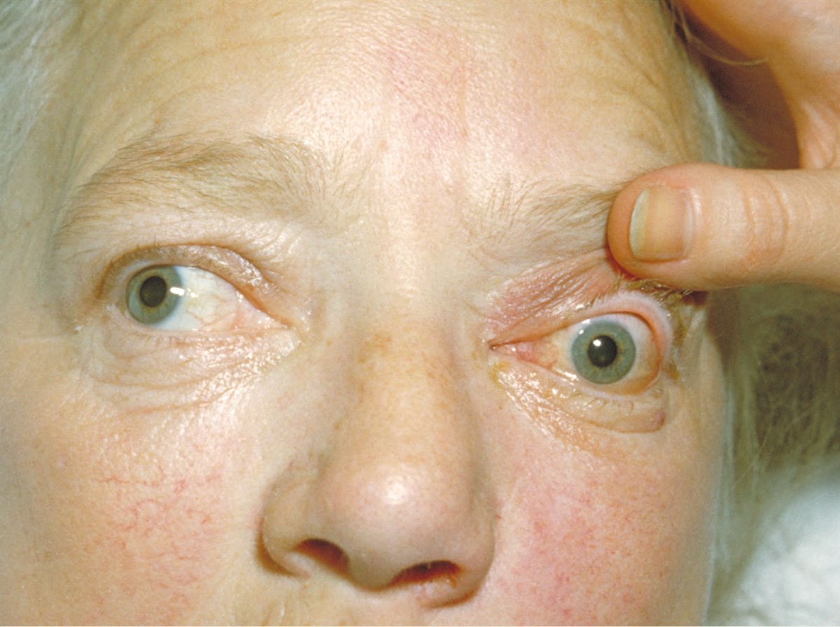
Proptosis — forward displacement of the globe out of the orbit — is a physical sign with a wide differential diagnosis.
The diagnostic approach to a proptotic eye is fundamentally different from the approach to almost any other ocular finding, because the orbit is a closed bony compartment: anything that adds volume within that space — inflammation, tumor, vascular malformation, hemorrhage, or fat — has one place to go, forward, pushing the globe out.
Thyroid eye disease is by far the most common cause in adults and already has its own detailed coverage on this site; this article focuses on building the broader differential and knowing when proptosis is not thyroid-related, since defaulting to that assumption in every case risks missing a genuinely different and sometimes more urgent underlying process.
Framework for the Differential
Laterality is the first and most useful discriminator: bilateral proptosis strongly favors thyroid eye disease, while unilateral proptosis broadens the differential considerably toward orbital tumors, vascular lesions, or inflammatory or infectious processes localized to one side.
Time course is the second key variable — acute onset over hours to days points toward infection, hemorrhage, or inflammation, while gradual progression over months points toward a slow-growing tumor or vascular malformation.
Pain is the third discriminator worth building into the initial assessment: painful proptosis suggests infection, inflammation (including thyroid eye disease in its active phase), or an acutely expanding vascular lesion, while painless proptosis is more typical of a slow-growing benign tumor.
Combining these three variables — laterality, tempo, and pain — before ever ordering imaging often narrows the likely category enough to guide which specific imaging study and how urgently it should be obtained, rather than defaulting to the same broad workup for every patient presenting with a protruding eye.
Major Causes by Category
- Thyroid eye disease — bilateral (though can be asymmetric), associated with lid retraction, restrictive myopathy, and other signs of Graves’ orbitopathy
- Orbital cellulitis — acute, painful, with fever and often a sinus source; a true emergency given the risk of vision loss and intracranial spread
- Orbital tumors — cavernous hemangioma (the most common benign orbital tumor in adults), lymphoma, lacrimal gland tumors, metastases, and, in children, rhabdomyosarcoma
- Vascular lesions — carotid-cavernous fistula (pulsatile proptosis with an audible bruit), orbital varix (proptosis that worsens with Valsalva or head-down positioning)
- Idiopathic orbital inflammation (orbital pseudotumor) — acute or subacute pain and proptosis without a clear infectious or neoplastic cause, often responsive to corticosteroids
- Trauma — retrobulbar hemorrhage, orbital emphysema, foreign body
- Pediatric-specific causes — rhabdomyosarcoma, neuroblastoma metastasis, and orbital cellulitis, all of which require urgent evaluation in a child
All-fit Slit-Lamp Adapter
Record and share exactly what you see at the slit lamp. One adapter fits any slit lamp or surgical microscope — and any smartphone.
From Choroida — the team behind this siteKey Exam Findings
Exophthalmometry quantifies the degree of proptosis and, importantly, allows objective tracking over time; a single measurement matters less than the trend on serial visits.
Palpation for a mass, auscultation for a bruit (suggesting a vascular lesion such as carotid-cavernous fistula), and assessment for resistance to retropulsion (globe pushed gently back into the orbit) all add useful information at the bedside before any imaging is obtained.
Visual acuity, color vision, and pupillary assessment for a relative afferent pupillary defect are essential in every case, because compressive optic neuropathy from any orbital process — not just thyroid eye disease — is the finding that most urgently changes management.
Extraocular motility should be checked for restriction, which points toward either infiltrative/inflammatory involvement of the muscles themselves or mechanical impingement from an adjacent mass.
Documenting these findings carefully at the initial visit, even when imaging is being arranged urgently, establishes a baseline against which the effect of treatment — or the significance of further progression before treatment begins — can later be judged.
Red Flags Requiring Urgent Evaluation
- Rapid progression over hours to days
- Fever or signs of systemic infection
- Reduced visual acuity, color vision, or an afferent pupillary defect
- Severe pain, particularly with eye movement
- Pulsatile proptosis or an audible bruit
Any of these findings warrants same-day imaging and, typically, same-day ophthalmology or emergency evaluation, because the underlying causes at this end of the spectrum — orbital cellulitis with abscess, retrobulbar hemorrhage, compressive optic neuropathy — can threaten vision within hours if not addressed, leaving little margin for a delayed or deferred workup.
Imaging
CT is typically the first-line study for acute proptosis, when trauma, sinus disease, or a suspected foreign body are in the differential, because it images bone well and is fast to obtain.
MRI is preferred when a soft-tissue mass, vascular lesion, or optic nerve involvement needs more detailed characterization, and is generally the study of choice for a more indolent, gradually progressive presentation where malignancy or a vascular malformation is being considered, given its superior soft-tissue resolution compared with CT.
Management
Management follows directly from the identified cause rather than the proptosis itself: systemic antibiotics (and often surgical drainage) for orbital cellulitis, corticosteroids for active thyroid eye disease or idiopathic orbital inflammation, and biopsy or excision for a suspected tumor.
Emergent lateral canthotomy and cantholysis is a sight-saving procedure reserved specifically for compartment syndrome from retrobulbar hemorrhage with optic nerve compression, and recognizing that specific scenario quickly is one of the highest-yield skills in orbital emergency care, since performing it promptly can be the difference between preserved and permanently lost vision.


Document what you see
Two smartphone imaging tools built for everyday clinic use — one for the slit lamp, one for the fundus.
From Choroida — the team behind this siteReferences
- Rootman J, ed. Diseases of the Orbit: A Multidisciplinary Approach.
- American Academy of Ophthalmology. Basic and Clinical Science Course, Section 7: Orbit, Eyelids, and Lacrimal System.
- Chaudhry IA, Shamsi FA, Elzaridi E, et al. Outcome of treated orbital cellulitis in a tertiary eye care center. Ophthalmology.
- Bagheri N, Wajda B, eds. The Wills Eye Manual.
Test yourself
A few questions straight from this article.
-
Why does any added orbital volume displace the globe forward in proptosis?
Inflammation, tumour, vascular malformation, haemorrhage or fat added to a closed bony orbit has only one direction to expand, pushing the globe forward. -
Bilateral proptosis in an adult most strongly favours which underlying cause?
Laterality is the first discriminator: bilateral proptosis strongly favours thyroid eye disease, while unilateral proptosis broadens the differential toward tumours, vascular lesions and localized inflammation. -
Proptosis progressing gradually over months without pain most suggests which category?
Gradual progression over months points to a slow-growing tumour or vascular malformation, and painless proptosis is typical of a slow-growing benign tumour. -
Which is the most common benign orbital tumour in adults?
The article names cavernous haemangioma as the most common benign orbital tumour in adults, among orbital causes that also include lymphoma, lacrimal gland tumours and metastases. -
Pulsatile proptosis with an audible orbital bruit points to which lesion?
Pulsatile proptosis with an audible bruit is the classic presentation of a carotid-cavernous fistula, and auscultation at the bedside can raise it before imaging. -
Proptosis that worsens with Valsalva or head-down positioning suggests which lesion?
An orbital varix characteristically produces proptosis that worsens with Valsalva or head-down positioning, distinguishing it from other vascular causes. -
Which orbital malignancy is a specifically paediatric cause of proptosis?
Paediatric-specific causes are rhabdomyosarcoma, neuroblastoma metastasis and orbital cellulitis, all of which require urgent evaluation in a child. -
What makes exophthalmometry most valuable in assessing a proptotic eye?
Exophthalmometry quantifies proptosis for objective follow-up; a single measurement matters far less than the trend documented on serial visits. -
Which imaging study is preferred for gradually progressive proptosis with a suspected orbital mass?
MRI better characterises soft-tissue masses, vascular lesions and optic nerve involvement, while CT is first-line in acute cases with trauma, sinus disease or foreign body. -
Emergent lateral canthotomy and cantholysis is reserved for which specific scenario?
Canthotomy with cantholysis is a sight-saving decompression for orbital compartment syndrome from retrobulbar haemorrhage with optic nerve compression, and recognising it quickly preserves vision.
