Bruch’s membrane is a thin, five-layer sheet between the retinal pigment epithelium and the choriocapillaris.
It supports exchange of nutrients and waste between the retina and the choroid.
Therefore, changes in this membrane sit at the root of several retinal diseases.
Structure of Bruch’s Membrane
Five layers make up the membrane.
They are the basement membrane of the retinal pigment epithelium, the inner collagenous layer, the elastic layer, the outer collagenous layer and the basement membrane of the choriocapillaris.
Also, the layers differ in composition and function.
Because the membrane is thin, it is easy to overlook on imaging.
Function of Bruch’s Membrane
The membrane acts as a filter and a barrier.
Nutrients, oxygen and retinoids pass across it toward the retina.
Also, waste products pass back toward the choroid.
Therefore, its permeability affects the health of the outer retina.
Fundus Explorer Pro
Photograph the retinal findings described here with the phone already in your pocket — 22 D optics and built-in illumination in one handheld unit.
From Choroida — the team behind this siteAging of Bruch’s Membrane
The membrane thickens with age.
Lipids accumulate, and collagen cross-links increase.
Also, the hydraulic conductivity falls, which slows the movement of fluid.
Because the membrane becomes a poor filter, waste products pile up in the outer retina.
Lipid Deposition in Bruch’s Membrane
Esterified and unesterified cholesterol accumulate in the inner collagenous layer.
Curcio and colleagues described a lipid wall that forms with age.
Also, this lipid-rich layer may act as a barrier to transport.
Therefore, it plays a role in early age-related macular degeneration.
Basal Deposits and Drusen
Basal laminar deposit lies between the plasma membrane of the RPE and its basement membrane.
Basal linear deposit lies within the inner collagenous layer.
Curcio and Millican found that basal linear deposit and large drusen are specific for early macular degeneration.
Also, drusen sit between the RPE basement membrane and the inner collagenous layer (see basal laminar drusen).
Bruch’s Membrane in Macular Degeneration
Thickening, lipid and calcification accompany disease.
Breaks allow choroidal vessels to grow into the sub-RPE or subretinal space.
Also, geographic atrophy follows loss of the RPE and the choriocapillaris (see age-related macular degeneration).
Because the membrane limits repair, damage can be permanent.
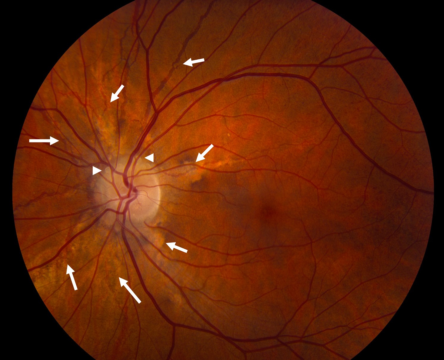
Angioid Streaks
Angioid streaks are breaks in a calcified and brittle membrane.
Pseudoxanthoma elasticum is the classic cause (see angioid streaks).
Also, other diseases include sickle cell disease and Paget disease.
Therefore, systemic evaluation is required when you see streaks.
Myopia and Lacquer Cracks
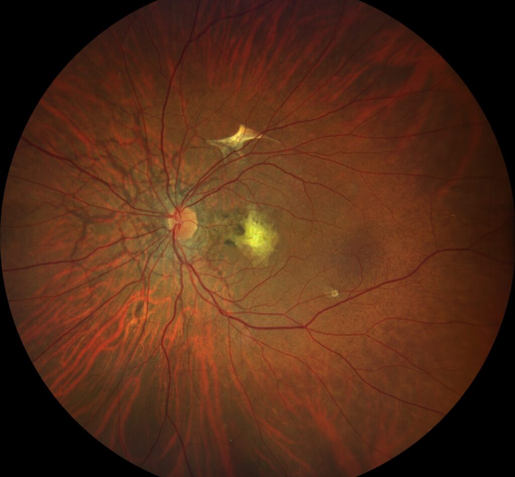
In pathologic myopia, the membrane stretches and breaks.
These breaks are called lacquer cracks.
Also, choroidal neovascularization can follow (see myopic choroidal neovascularization).
Because the eye is long, mechanical stress drives the damage.
Genetic Disorders
Sorsby fundus dystrophy involves the deposition of abnormal TIMP3 in the membrane.
Doyne honeycomb dystrophy involves drusen-like deposits.
Also, these conditions show that the membrane is a target of inherited disease (see Sorsby fundus dystrophy).
Imaging Bruch’s Membrane
Optical coherence tomography shows the RPE and Bruch complex as one band.
Separation between them marks a pigment epithelial detachment (see pigment epithelial detachment).
Also, double-layer sign suggests a neovascular membrane.
Therapeutic Ideas
Researchers study ways to improve transport across the membrane.
Approaches include reducing lipid and restoring permeability.
Also, tissue engineering aims to replace damaged membranes.
Because these ideas are early, they remain experimental.
Clinical Summary
Think of Bruch’s membrane when you see drusen, streaks, cracks or breaks.
Also, remember that a healthy membrane is vital for the RPE.
Hydraulic Conductivity and Fluid Movement
Fluid moves from the retina toward the choroid across the membrane.
With age, the movement slows.
Also, this change may contribute to fluid accumulation in disease.
Because it is hard to measure in life, most data come from donor eyes.
Calcification and Fragility
Calcium deposits make the membrane brittle.
Mechanical stress then causes breaks.
Also, the process is more rapid in pseudoxanthoma elasticum.
Therefore, patients may need regular follow-up.
Choroidal Neovascularization and Breaks
New vessels pass through breaks in the membrane.
Treatment targets the growth factors that drive them.
Also, the break remains and may allow recurrence.
Because the membrane does not heal well, recurrence is common.
Relevance for Surgery
Surgeons see the membrane after removal of a neovascular complex.
Also, subretinal surgery requires caution not to damage it.
Therefore, gentle manipulation helps protect the RPE.
For the latest studies, see the PubMed results for Bruch’s membrane.


Document what you see
Two smartphone imaging tools built for everyday clinic use — one for the slit lamp, one for the fundus.
From Choroida — the team behind this siteReferences
- Curcio CA, Millican CL. Basal linear deposit and large drusen are specific for early age-related maculopathy. Arch Ophthalmol. 1999;117:329-339.
- Booij JC, Baas DC, Beisekeeva J, Gorgels TG, Bergen AA. The dynamic nature of Bruch’s membrane. Prog Retin Eye Res. 2010;29:1-18.
- Bhutto I, Lutty G. Understanding age-related macular degeneration (AMD): relationships between the photoreceptor/retinal pigment epithelium/Bruch’s membrane/choriocapillaris complex. Mol Aspects Med. 2012;33:295-317.
- Spraul CW, Lang GE, Grossniklaus HE, Lang GK. Histologic and morphometric analysis of the choroid, Bruch’s membrane, and retinal pigment epithelium in postmortem eyes with age-related macular degeneration and histologic examination of surgically excised choroidal neovascular membranes. Surv Ophthalmol. 1999;44(Suppl 1):S10-S32.

Writes and reviews the clinical articles on Educate. Every post is checked against peer-reviewed sources before it goes live.