
CASE REPORT
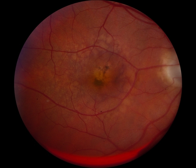
A 55-year-old female visited an eye hospital for her routine eye checkup with a complaint of decreased near vision. On history taking, she was found diabetic as well as hypertensive.
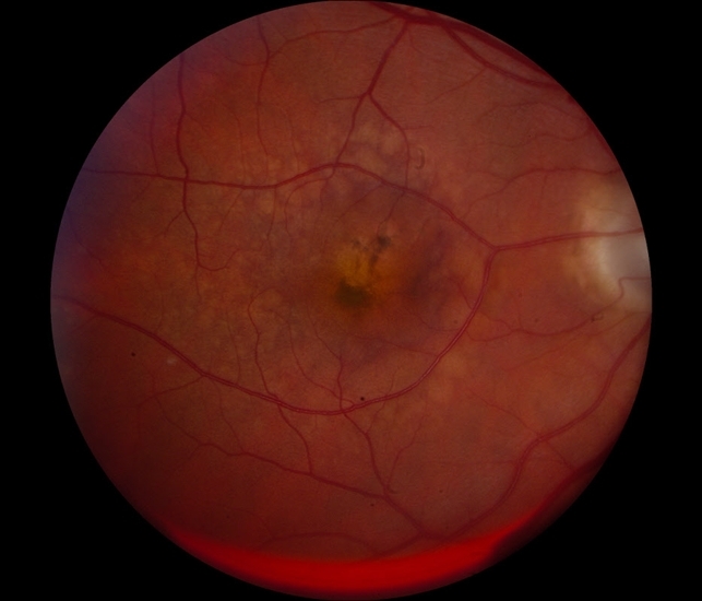
Her best corrected visual acuity was 6/6 in both eyes. Adnexa was normal in both eyes. Bilateral Pupil reaction was Round Regular Reactive (RRR). Bilateral media was clear.
On slit lamp examination, the optic disc, retinal vasculature and macula were found normal but an extramacular discoloured area temporal to the macula was noted.
The patient was kept on follow-up to observe fundus changes and visual outcomes after some time. Followup reports also confirm the diagnosis of Pigment Epithelial detachment (PED).
Pigment Epithelial detachment (PED) entity
Pigment Epithelial detachment (PED) refers to a detachment of Retinal Pigment Epithelium from the Bruch’s membrane. This detachment creates a space between RPE and Bruch’s membrane which is occupied by blood, drusenoid materials and exudates.
PEDs can be caused by chorioretinal diseases such as Age-related macular degeneration (ARMD), Central serous Chorioretinopathy(CSC), and polypoidal choroidal vasculopathy (PCV).
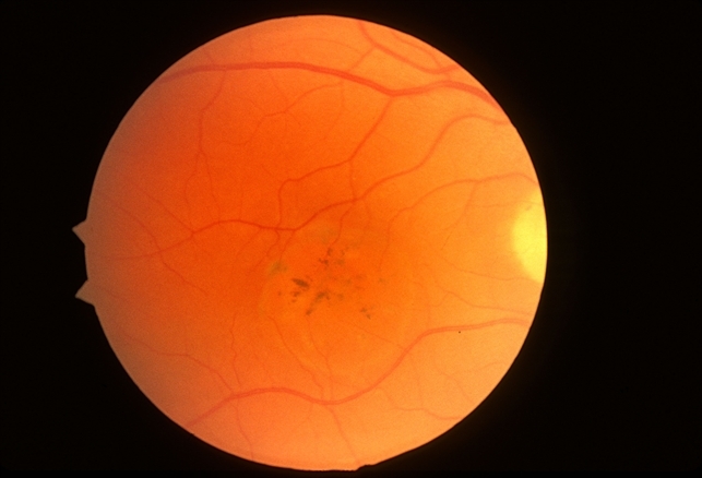
Types of PED:
In AMD, PED can be categorized into the following main types:
1. Serous PED
2. Drusenoid PED
3. Fibrovascular PED
4. Haemorrhagic PED
1. Serous PED:
- Patients of serous PED present with the symptoms of painless blurring of vision, micropsia, Metamorphopsia and distortion of objects. They might have developed induced hyperopia.
- On slit lamp, it is characterized by yellow, orange, dome-shaped elevations of RPE having distinct margins.
- Fluid beneath the RPE shows transillumination when examined through a slit lamp.
- On OCT, PEDs are seen as elevations of RPE with a hyporeflective sub-RPE space.
- OCT is performed to differentiate whether PED is caused by AMD or CSC. If it’s due to AMD, subfoveal Choroidal thickness appears normal. Subfoveal Choroidal thickness will be increased in case of CSC.
- On FA, this type of PED shows early hyperfluorescence and pooling without showing leakage.
2. Drusenoid PED:
- It is characterized by irregular surface yellow white Elevations of RPE with scalloped borders present within the macular region.
- It is mostly bilateral.
- FA indicates early mild hyperfluorescence with late staining without showing any leakage.
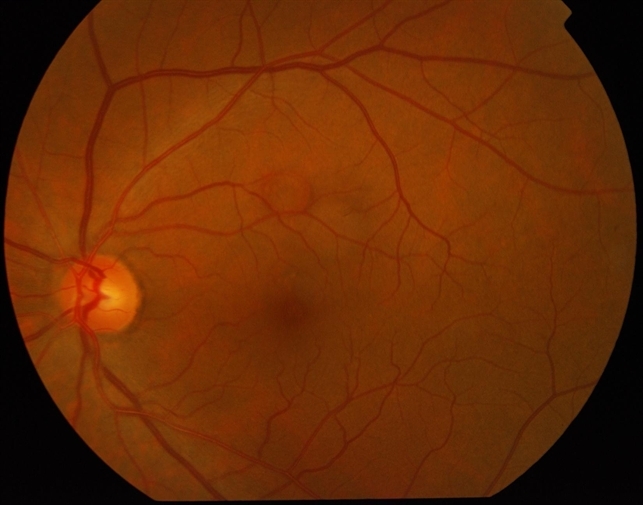
- Fibrovascular PED:
- It occurs due to the presence of CNV. Due to breaks in Bruch’s membrane, abnormal blood vessels from the choroid grow beneath the RPE. These abnormal vessels lack a blood-retinal barrier, so they leak and bleed beneath the RPE, separating it from Bruch’s membrane and leading to Fibrovascular PED.
- It is characterized by irregular surface RPE elevations with haemorrhages, intra-retinal fluid and subretinal fluid etc
- FA shows stippled hyperfluorescence with late leakage.
- Haemorrhagic PED:
- This type of PED is associated with CNV or Polypoidal Choroidal Vasculopathy (PCV).
- Patients with Haemorrhagic PED present with sudden Impairment of central vision.
- On examination, dark red dome-shaped elevated lesions with sharp borders are noted.
- FA shows masking of background fluorescence, but overlying vessels are visible.
Fundus Explorer Pro
Photograph the retinal findings described here with the phone already in your pocket — 22 D optics and built-in illumination in one handheld unit.
From Choroida — the team behind this siteMANAGEMENT of Pigment Epithelial detachment (PED)
- For serous PED, no treatment option is available.
- For Vascularized PED, Laser photocoagulation, anti-VEGF therapy, photodynamic therapy and intravitreal steroids can be given as a treatment.


Document what you see
Two smartphone imaging tools built for everyday clinic use — one for the slit lamp, one for the fundus.
From Choroida — the team behind this siteREFERENCES
- American Academy of Ophthalmology. Pigment epithelial detachment.
- Khochtali S, Ksiaa I, Megzari K, Khairallah M. Retinal Pigment Epithelium Detachment in Acute Vogt-Koyanagi-Harada Disease: An Unusual Finding at Presentation. Ocul Immunol Inflamm. 2019;27(4):591–594.
- Mrejen, S. (2013). Multimodal imaging of pigment epithelial detachment: a guide to evaluation. Retina, 33(9), 1735-1762.
-
Gass JD, Norton EW, Justice J., Jr Serous detachment of the retinal pigment epithelium. Trans Am Acad Ophthalmol Otolaryngol. 1966;70:990–1015.
-
Gass JD. Drusen and disciform macular detachment and degeneration. Trans Am Ophthalmol Soc. 1972;70:409–436.
-
Casswell AG, Kohen D, Bird AC. Retinal pigment epithelial detachments in the elderly: classification and outcome. Br J Ophthalmol. 1985;69:397–403.
Test yourself
A few questions straight from this article.
-
What is a pigment epithelial detachment (PED)?
PED is detachment of the retinal pigment epithelium from Bruch's membrane, leaving a space between them that can hold blood, drusenoid material and exudate. -
Which chorioretinal disease is listed as a cause of pigment epithelial detachment?
PED can be caused by chorioretinal diseases such as age-related macular degeneration, central serous chorioretinopathy and polypoidal choroidal vasculopathy. -
Which refractive change may a patient with a serous pigment epithelial detachment develop?
Serous PED causes painless blurring, micropsia, metamorphopsia and distortion, and the patient may develop induced hyperopia. -
How does a serous pigment epithelial detachment appear on slit-lamp fundus examination?
Serous PED is seen as a yellow-orange, dome-shaped elevation of the RPE with distinct margins; scalloped borders belong to drusenoid PED and a dark red dome to haemorrhagic PED. -
On OCT, which finding favours central serous chorioretinopathy over AMD as the cause of a PED?
OCT helps find the cause of a PED: subfoveal choroidal thickness is increased in CSC but appears normal when the PED is due to AMD. -
Which fluorescein angiography pattern identifies a fibrovascular pigment epithelial detachment?
Fibrovascular PED shows stippled hyperfluorescence with late leakage, unlike serous PED (pooling without leakage), drusenoid PED (late staining) and haemorrhagic PED (masking). -
Which laterality is typical of drusenoid pigment epithelial detachment?
Drusenoid PED is mostly bilateral and appears as irregular yellow-white RPE elevations with scalloped borders within the macula. -
How do patients with a haemorrhagic pigment epithelial detachment typically present?
Haemorrhagic PED, associated with CNV or polypoidal choroidal vasculopathy, presents with sudden impairment of central vision and a dark red dome-shaped elevation. -
Which process underlies a fibrovascular pigment epithelial detachment?
Fibrovascular PED occurs due to the presence of CNV and shows irregular RPE elevation with haemorrhage, intraretinal fluid and subretinal fluid. -
Which treatments are listed for a vascularised pigment epithelial detachment?
Vascularised PED can be treated with laser photocoagulation, anti-VEGF therapy, photodynamic therapy and intravitreal steroids.
