

CASE REPORT
A 72-year-old retired school teacher presented to an eye clinic with a six-month history of gradual vision loss in her left eye, expressing difficulty in recognizing faces, reading, and engaging in daily activities requiring detailed vision.
Her medical history includes controlled hypertension and hypercholesterolemia managed with statins, with no history of diabetes or smoking.
The patient reported a familial predisposition to vision loss, as her mother experienced a similar condition in later years.
Throughout her life, she maintained regular eye examinations with an optometrist and had no significant ocular surgeries or injuries, wearing glasses for mild myopia and presbyopia.
Clinical examination revealed a visual acuity of 20/25 in the right eye and 20/200 in the left, with Amsler grid distortion and a central scotoma.
Smartphone Fundus examination displayed drusen and pigmentary changes in the left macula, while OCT confirmed subretinal fluid and atrophy.
Fluorescein angiography revealed choroidal neovascularization, leading to the diagnosis of Age-Related Macular Degeneration (AMD).
DISEASE
Age-related macular degeneration (ARMD) is an acquired degeneration of the retina that causes significant central visual impairment through a combination of non-neovascular (drusen and retinal pigment epithelium abnormalities), and neovascular derangement (choroidal neovascular membrane formation).
Advanced disease may involve focal areas of retinal pigment epithelium (RPE) loss, subretinal or sub-RPE hemorrhage or serous fluid, as well as subretinal fibrosis.
A genetic underpinning is inferred from its predilection to those of European ancestry, although environmental, nutritional, and developmental (ie., aging) processes interact to affect the degeneration observed in the macula.
Newly implicated biochemical pathways combined with a paucity of treatment options for the majority of ARMD (i.e., dry ARMD) have created fertile ground for novel therapeutics.
Fundus Explorer Pro
Photograph the retinal findings described here with the phone already in your pocket — 22 D optics and built-in illumination in one handheld unit.
From Choroida — the team behind this siteClassification
- Early ARMD: Defined by the presence of numerous small (<63 microns, “hard”) or intermediate (≥63 microns but <125 microns, “soft”) drusen. Note: Small drusen are frequently seen in those 50 and older, and can represent an epiphenomenon of aging (therefore, intermediate drusen are more specific for ARMD).
- Intermediate ARMD: Macular disease characterized by either extensive drusen of small or intermediate size or any drusen of large size (≥125 microns). 124 microns is the average diameter of the retinal vein at the optic disc margin.
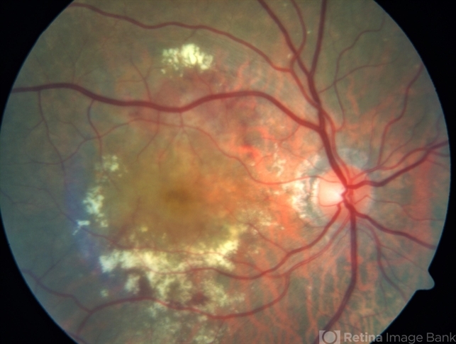
- Advanced ARMD: Defined by the presence of either geographic atrophy or choroidal neovascular membrane (along with its sequelae, such as subretinal or sub-RPE hemorrhage or serous fluid, and subretinal fibrosis). The figure below demonstrates progressive geographic atrophy over a 29-month period. There is also an area of choroidal neovascularization along the inferior aspect of the scar with subretinal fibrosis that progresses over these 29 months.
Etiology
A combination of risk factors interplay to modify the Bruch’s membrane/choroid complex, the retinal pigment epithelium, and photoreceptor cells.
The initiating events affect one, both, or all of these tissue components. A change in one of these tissue components is thought to impart an influence on the others in such a way that an ‘intermediate disease mechanism” arises.
The degenerating retina succumbs to the final endpoint of geographic atrophy, choroidal neovascularization, and pigment epithelial detachment.
Treatments targeting intermediate disease mechanisms or initiating disease factors are in the minority but may offer a more successful approach to vision preservation than those targeting relatively later steps in ARMD pathophysiology (i.e., choroidal neovascularization).
Risk Factors

Age
ARMD risk increases with age. The risk increases more than three-fold in patients older than 75 years of age compared to the group of patients between 65-74 years of age.
Cigarette Smoking
A ten-pack-year tobacco smoking history is associated with increased development of exudative age-related macular degeneration.
After controlling for confounding factors such as socioeconomic status, alcohol consumption, and cardiovascular disease, the rates of visually significant ARMD in smokers and non-smokers over the age of 75 were calculated in a United Kingdom population-based cross-sectional study.
Current smokers were twice as likely to have ARMD-related vision loss when compared to non-smokers, ex-smokers had a slightly increased risk (odds ratio of 1.13), and those who had stopped smoking over 20 years earlier were not at increased risk for developing vision loss from ARMD.
Genetic susceptibility
In addition to increasing one’s risk of developing ARMD, certain genetic loci have been associated with variable effects on treatment response, such as intravitreal anti-VEGF agents.
Complement factor H (CFH) is an important gene in the pathogenesis of ARMD.
Pharmacogenomic studies, such as these, may guide treatment regimens in the future, or may at least provide a more accurate prediction of treatment response.
Recently, microRNA dysregulation has been linked with the development of ARMD, modulation of which could provide potential treatments for the disease.
Other Risk factors
Cardiovascular disease, Hypertension, Female gender, White race, Hypercholesterolemia, Obesity, Hyperopia, Family History, Light Irides.
MANAGEMENT
Observation with risk factor modification and nutritional supplementation is the mainstay of treatment for nonexudative ARMD.
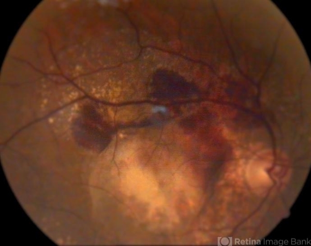
However, current studies are underway to evaluate complement pathway inhibition for the treatment of geographic atrophy in patients with nonexudative ARMD though no treatment modality is currently approved by the FDA for the treatment of geographic atrophy.
Exudative ARMD is managed with more closely spaced examinations and intravitreal injections of anti-VEGF agents or laser treatments.
Read more about management strategies
Patients with advanced disease in both eyes should undergo evaluation and rehabilitation with low-vision services.


Document what you see
Two smartphone imaging tools built for everyday clinic use — one for the slit lamp, one for the fundus.
From Choroida — the team behind this siteREFERENCES
- Bressler NM, Bressler SB, Sarks SH, Sarks JP. Chapter 60. Age-Related Macular Degeneration: Nonneovascular Early AMD, Intermediate AMD, and Geographic Atrophy, Volume II, 4th Edition. Elsevier, Mosby. 2006. Editor: Andrew SP. Schachat.
- JRW Yates, T Sepp, BK Matharu, JC Khan, DA Thurlby, H Shahid, DG Clayton, C Hayward, J Morgan, AF Wright, AM Armbrecht, B Dhillon, IJ Deary, E Redmond, AC Bird, AT Moore, The Genetic Factors in AMD Study Group. Complement C3 variant and the risk of age-related macular degeneration. N. Engl. J. Med. 357:553-561 (2007).
- ↑ GS Hageman, DH Anderson, LV Johnson, LS Hancox, AJ Taiber, LI Hardisty, JL Hageman, HA Stockman, JD Borchardt, KM Gehrs, RJ Smith, G Silvestri, SR Russell, CC Klaver, I Barbazetto, S Chang, LA Yannuzzi, GR Barile, et al. A common haplotype in the complement regulatory gene factor H (HF1/CFH) predisposes individuals to age-related macular degeneration. Proc. Natl. Acad. Sci. USA 20:7227-7232 (2005)
- JL Haines, MA Hauser, S Schmidt, WK Scott, LM Olson, P Gallins, KL Spencer, SY Kwan, M Noureddine, JR Gilbert, N Schnetz-Boutaud, A Agarwal, EA Postel, MA Pericak-Vance. The complement factor H variant increases the risk of age-related macular degeneration. Science 308:419-421 (2005).
- RJ Klein, C Zeiss, EY Chew, J-Y Tsai, RS Sackler, C Haynes, AK Henning, JP SanGiovanni, SM Mane, ST Mayne, MB Bracken, FL Ferris, J Ott, C Barnstable, J Hoh. Complement factor H polymorphism in age-related macular degeneration. Science 308:385-389 (2005).
- AO Edwards, R Ritter III, KJ Abel, A Manning, C Panhuysen, LA Farrer. Complement factor H polymorphism and age-related macular degeneration. Science 308:421-424 (2005).
Test yourself
A few questions straight from this article.
-
Age-related macular degeneration is best described as which kind of disorder?
ARMD is an acquired degeneration of the retina causing significant central visual impairment, through non-neovascular changes (drusen and RPE abnormality) and neovascular derangement. -
In the classification of age-related macular degeneration, drusen are called large at what size?
Large drusen measure 125 microns or more. Small drusen are under 63 microns and intermediate drusen are 63 microns or more but under 125 microns. -
Which finding defines intermediate age-related macular degeneration?
Intermediate ARMD is macular disease with either extensive drusen of small or intermediate size, or any drusen of large size. -
Why are intermediate drusen regarded as more specific for AMD than small drusen?
Small drusen are frequently seen in people aged 50 and older and can represent an epiphenomenon of ageing, so intermediate drusen carry more specificity for ARMD. -
What defines advanced age-related macular degeneration?
Advanced ARMD is defined by geographic atrophy or a choroidal neovascular membrane, together with sequelae such as subretinal or sub-RPE haemorrhage, serous fluid and subretinal fibrosis. -
Risk factors for age-related macular degeneration act on which tissue components to initiate the disease?
Risk factors interplay to modify the Bruch's membrane/choroid complex, the retinal pigment epithelium and the photoreceptors; change in one is thought to influence the others, generating an intermediate disease mechanism. -
How does AMD risk in patients older than 75 compare with patients aged 65 to 74?
ARMD risk rises with age, increasing more than three-fold in patients older than 75 compared with those aged 65 to 74. -
In the UK population study of smoking and AMD, which group over 75 had no increased risk of vision loss?
Current smokers were twice as likely to have ARMD-related vision loss, ex-smokers carried a slightly raised risk with an odds ratio of 1.13, and those who had stopped more than 20 years earlier were not at increased risk. -
Which gene is highlighted as important in the pathogenesis of age-related macular degeneration?
Complement factor H (CFH) is an important gene in ARMD pathogenesis, and certain genetic loci also affect the response to treatments such as intravitreal anti-VEGF agents. -
What is the mainstay of treatment for nonexudative age-related macular degeneration?
Nonexudative ARMD is managed by observation with risk factor modification and nutritional supplementation. Exudative disease instead needs closely spaced examinations with intravitreal anti-VEGF agents or laser.

Writes and reviews the clinical articles on Educate. Every post is checked against peer-reviewed sources before it goes live.


