
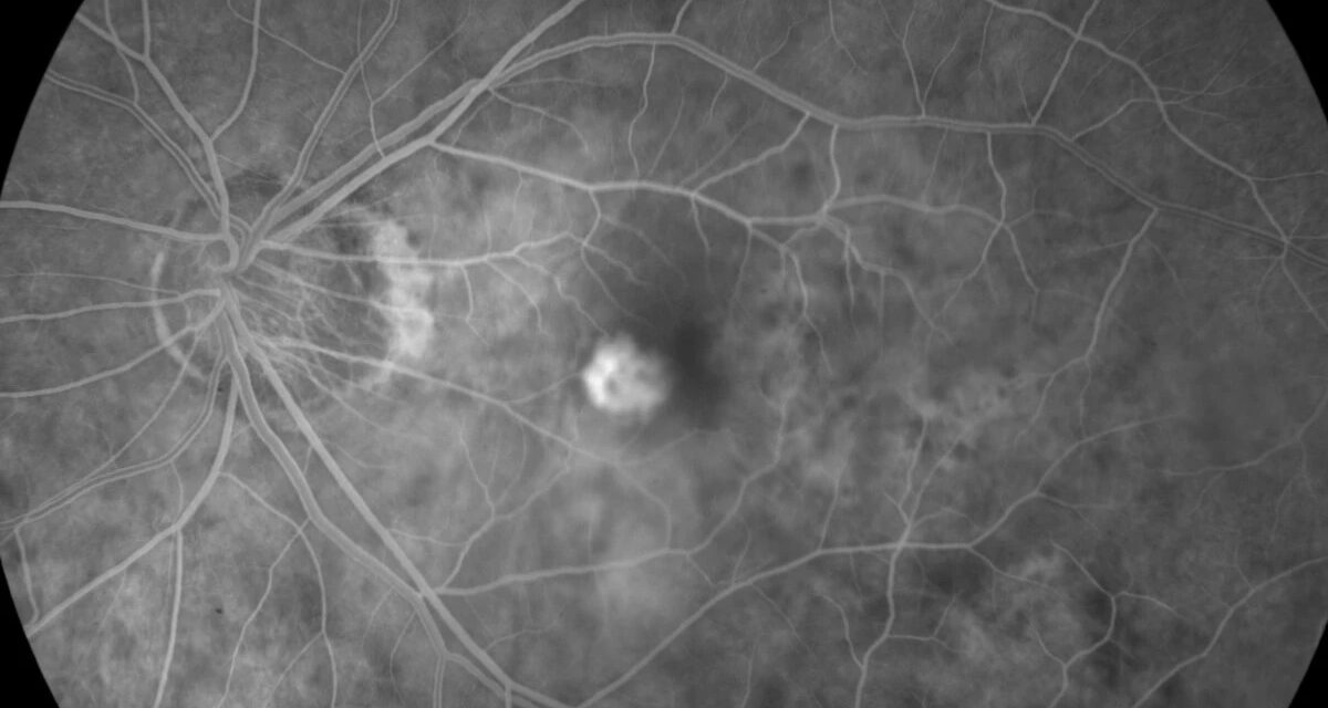
Myopic choroidal neovascularization (mCNV) is the most common vision-threatening complication of pathologic myopia, developing in a meaningful proportion of highly myopic eyes over a lifetime.
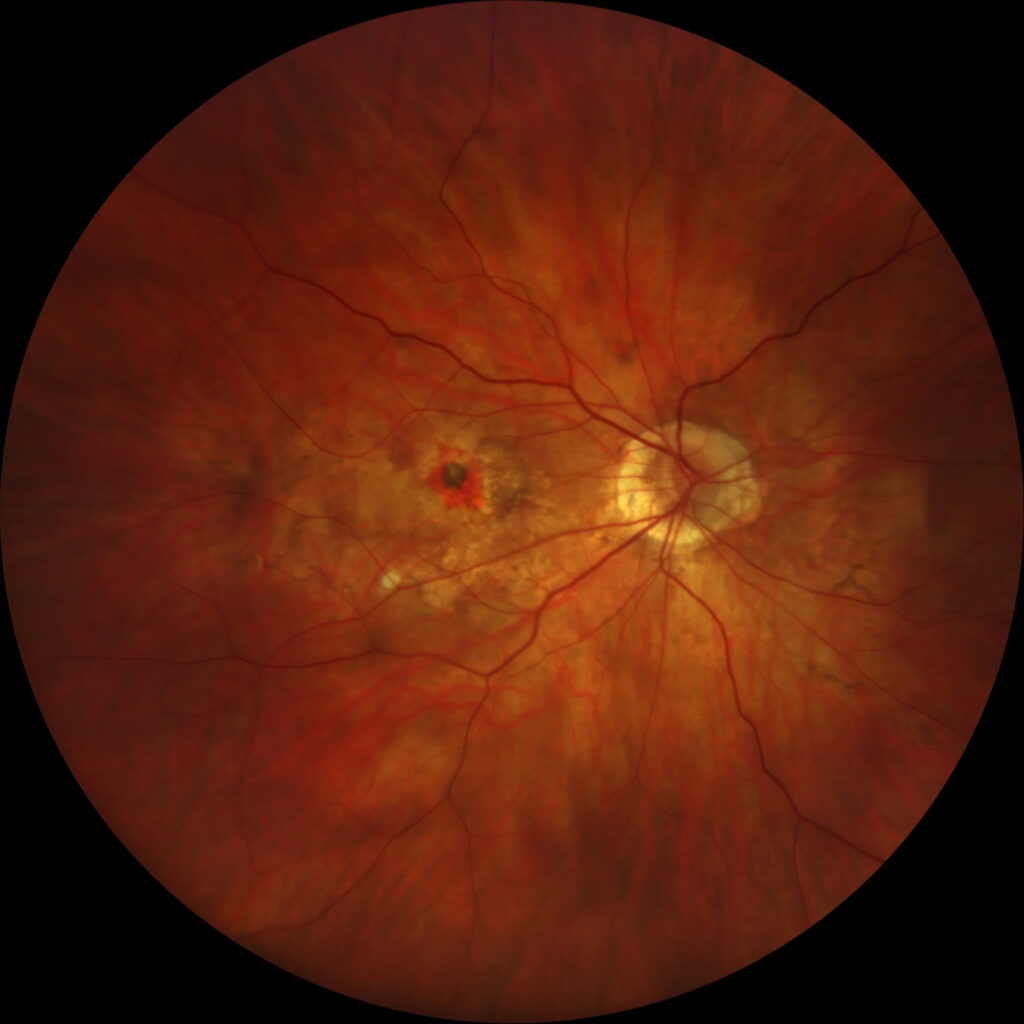
It is a different disease from age-related macular degeneration in almost every respect that matters for prognosis and treatment planning: younger patients, smaller lesions, less exudation, and, importantly, a better overall response to a shorter course of treatment than typical neovascular AMD.
Pathogenesis
Pathologic myopia produces progressive axial elongation of the globe, which stretches and thins the choroid, retinal pigment epithelium, and Bruch’s membrane over time.
Breaks in Bruch’s membrane — visible clinically as lacquer cracks — create a pathway for choroidal new vessels to grow through into the subretinal space.
MCNV frequently arises at or near the site of one of these breaks, distinguishing its mechanism from the drusen-driven RPE dysfunction that underlies typical age-related neovascular disease.
Because the driving process is mechanical stretching of an already thin, elongated eye rather than the age-related lipid and drusen accumulation of AMD, mCNV can develop in patients decades younger than the typical AMD population, and its natural history — smaller lesion size, less exudation, more limited growth potential — reflects this fundamentally different underlying biology.
Risk Factors
- High myopia, generally defined as greater than -6.00 diopters or axial length beyond 26 mm, with risk increasing further with more extreme myopia
- Presence of lacquer cracks on exam or imaging
- Posterior staphyloma
- Older age within the myopic population (though still generally younger rather than typical AMD patients)
- Fellow eye involvement — a history of mCNV in one eye significantly raises the risk of the second eye developing it as well
Fundus Explorer Pro
Photograph the retinal findings described here with the phone already in your pocket — 22 D optics and built-in illumination in one handheld unit.
From Choroida — the team behind this siteClinical Presentation
Patients report acute or subacute central vision loss, metamorphopsia, or a new central scotoma, developing against a background of longstanding high myopia and often against the additional visual limitation already present from myopic degenerative changes, which can make new symptoms harder for patients to characterize precisely.
Because the underlying myopic maculopathy already causes a degree of chronic visual limitation in many of these patients, a sudden change should be taken seriously rather than attributed to gradual progression of the underlying myopic degeneration.
Given how young many affected patients are relative to the typical AMD population, a sudden central visual change in a highly myopic patient in their thirties or forties should prompt the same urgency of evaluation as it would in an older patient with suspected AMD, rather than being dismissed simply because of the patient’s age.
Exam and Imaging Findings
- A small, grayish-green subretinal lesion, often with associated subretinal fluid or hemorrhage, typically smaller than the lesions seen in neovascular AMD
- Lacquer cracks — fine, yellowish, often branching lines in the posterior pole, representing breaks in Bruch’s membrane
- Background findings of myopic degeneration — posterior staphyloma, chorioretinal atrophy, tessellated fundus from visible choroidal vessels through a thinned RPE
- OCT showing a small, well-defined subretinal hyperreflective lesion, generally with less associated fluid than typical neovascular AMD
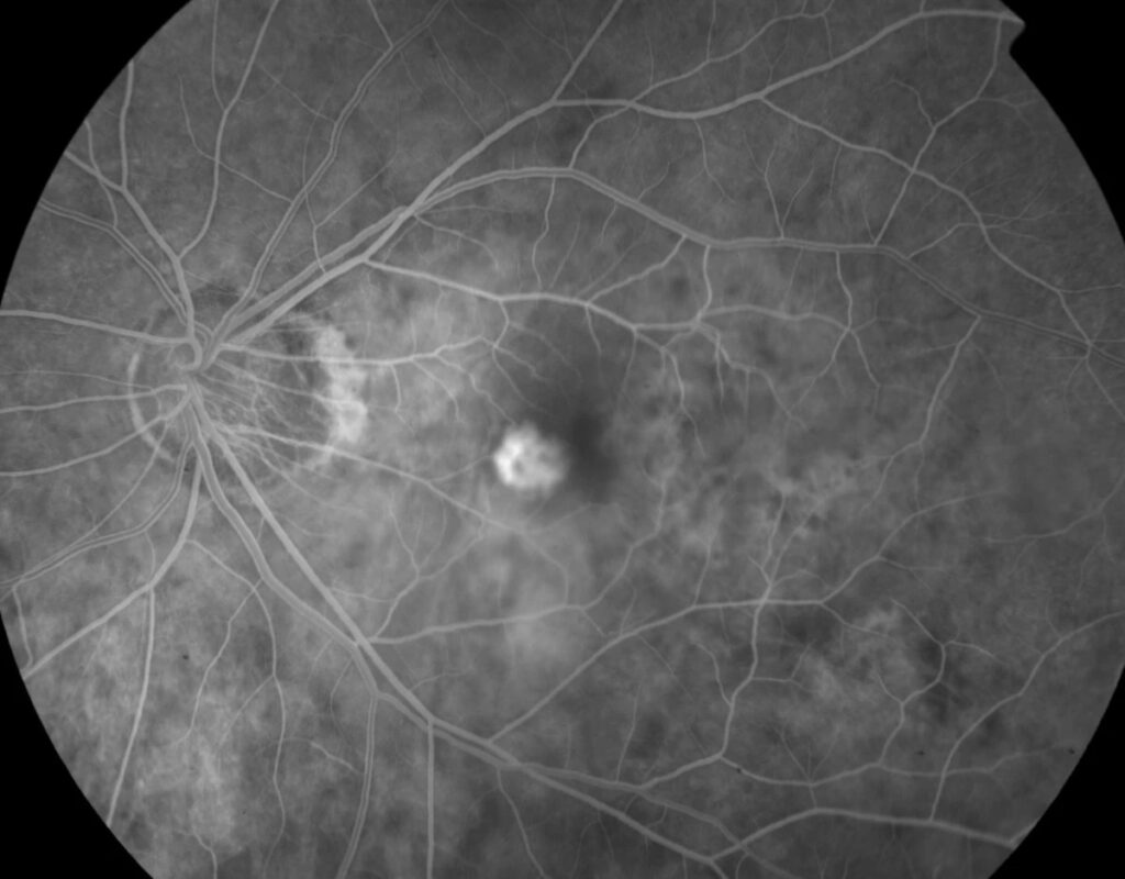
- OCT angiography and fluorescein angiography both useful for confirming the neovascular lesion and distinguishing it from other causes of subretinal change in a myopic eye
Differential Diagnosis
- Fuchs spot — an old, inactive, pigmented scar from prior mCNV, rather than an active lesion
- Simple subretinal hemorrhage without neovascularization, sometimes occurring after minor trauma or Valsalva in a highly myopic eye with fragile Bruch’s membrane
- Focal choroidal excavation or dome-shaped macula — structural myopic changes that can alter the macular contour without representing active neovascularization
- Age-related macular degeneration, in an older myopic patient where the two conditions can occasionally coexist
Management
Anti-VEGF therapy (ranibizumab, aflibercept, or bevacizumab) is first-line treatment and has transformed outcomes for mCNV, with many patients achieving good, durable visual results after a shorter treatment course than is typical for neovascular AMD.
A substantial proportion of eyes require only a few injections total, rather than the ongoing, often indefinite treatment schedule common in AMD, and this difference in expected treatment burden is worth explaining to patients up front, since it shapes expectations differently from what many patients may have heard about anti-VEGF therapy for AMD from family or friends.
Verteporfin photodynamic therapy was the standard treatment before anti-VEGF agents became available and is now used only rarely, generally reserved for cases where anti-VEGF therapy is not accessible or has not achieved adequate control.
Regular monitoring after apparent resolution remains important, since recurrence can occur, and the fellow eye should be monitored given the elevated risk of bilateral involvement over time.
Prognosis
Visual outcomes for mCNV, particularly when treatment is started promptly, are generally more favorable than for neovascular AMD, with many patients recovering meaningful vision and maintaining it over years with only occasional further treatment.
The long-term prognosis, however, is also shaped by the progression of the underlying myopic maculopathy itself, since the same axial elongation and choroidal thinning that predisposed the eye to mCNV in the first place continues to affect visual function independently of whether the neovascular lesion itself remains quiet, making ongoing, lifelong surveillance appropriate even in eyes with an apparently well-controlled, inactive lesion.


Document what you see
Two smartphone imaging tools built for everyday clinic use — one for the slit lamp, one for the fundus.
From Choroida — the team behind this siteReferences
- Wong TY, Ohno-Matsui K, Leveziel N, et al. Myopic choroidal neovascularisation: current concepts and update on clinical management. British Journal of Ophthalmology.
- Ikuno Y, Ohno-Matsui K, Wong TY, et al. Intravitreal aflibercept injection in patients with myopic choroidal neovascularization: the MYRROR study. Ophthalmology.
- Wolf S, Balciuniene VJ, Laganovska G, et al. RADIANCE: a randomized controlled study of ranibizumab in patients with choroidal neovascularization secondary to pathologic myopia. Ophthalmology.
- American Academy of Ophthalmology. Basic and Clinical Science Course, Section 12: Retina and Vitreous.
Test yourself
A few questions straight from this article.
-
What is the most common vision-threatening complication of pathologic myopia?
Myopic CNV is the leading vision-threatening complication of pathologic myopia and develops in a meaningful proportion of highly myopic eyes over a lifetime. -
Through which structural defect do new vessels reach the subretinal space in myopic CNV?
Axial elongation stretches and thins the choroid, RPE and Bruch's membrane; breaks in Bruch's membrane open a pathway, and the lesion often arises at or near one of them. -
How is the high myopia that predisposes to myopic CNV generally defined?
High myopia is generally taken as greater than -6.00 diopters or axial length beyond 26 mm, with risk rising further as myopia becomes more extreme. -
What does an active myopic CNV lesion look like on fundus examination?
Myopic CNV lesions are small and grayish-green with some subretinal fluid or hemorrhage, and are typically smaller and less exudative than neovascular AMD lesions. -
How do lacquer cracks appear in the myopic posterior pole?
Lacquer cracks are fine yellowish branching lines in the posterior pole that represent breaks in Bruch's membrane, and their presence is itself a risk factor for myopic CNV. -
What is a Fuchs spot in a highly myopic eye?
A Fuchs spot is the inactive pigmented scar left by a previous neovascular episode, not an active lesion needing treatment. -
How should sudden central visual change in a highly myopic 35-year-old be handled?
Many patients with myopic CNV are decades younger than the typical AMD population, so a sudden change deserves the same urgency and should not be dismissed on age grounds. -
What is first-line treatment for myopic choroidal neovascularization?
Ranibizumab, aflibercept or bevacizumab is first-line and has transformed outcomes, with many eyes needing only a few injections rather than the indefinite schedule typical of AMD. -
What is the current role of verteporfin photodynamic therapy in myopic CNV?
Photodynamic therapy was the standard before anti-VEGF agents and is now used only rarely, mainly when anti-VEGF is inaccessible or has not achieved adequate control. -
Why does an eye with a quiet, treated myopic CNV still need lifelong surveillance?
The axial elongation and choroidal thinning that caused the lesion continue to damage visual function independently, and the fellow eye also carries a raised risk.
