
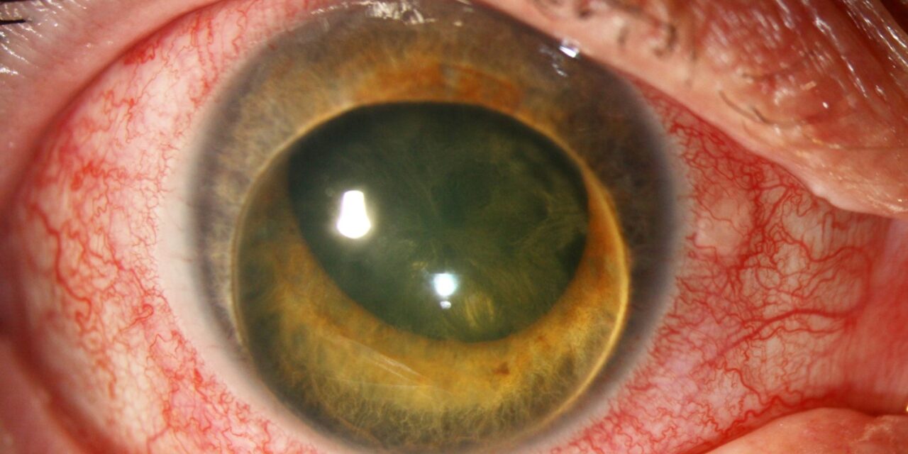
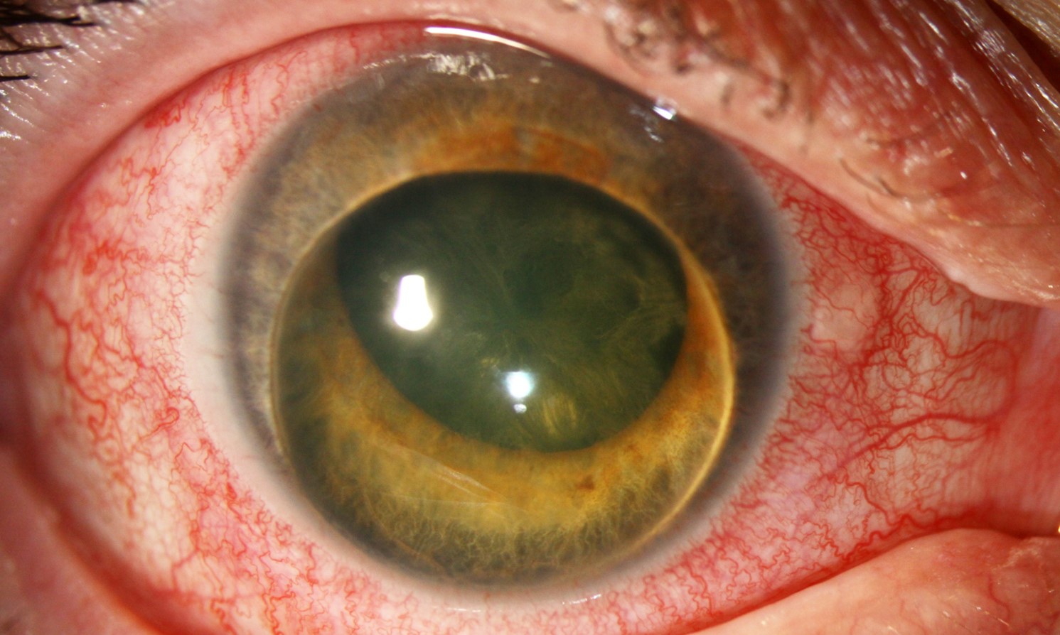
Phacomorphic glaucoma is acute angle closure caused directly by a swollen, intumescent cataractous lens pushing the iris forward and crowding the anterior chamber angle, and unlike primary angle closure, where the underlying anatomy predisposes to attack, this form develops because the lens itself has physically outgrown the space it once fit into, making cataract extraction, rather than the laser iridotomy used for primary angle closure, the treatment that actually addresses the cause.
How a Cataract Causes Angle Closure
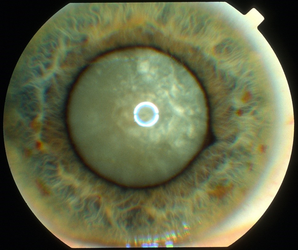
As a cataract matures, particularly in certain intumescent or morgagnian subtypes, the lens can absorb fluid and swell, increasing significantly in anteroposterior thickness.
This swollen lens pushes the iris-lens diaphragm forward, shallowing the anterior chamber and progressively crowding the trabecular meshwork at the angle, and when this crowding becomes severe enough to obstruct aqueous outflow, intraocular pressure rises acutely, producing a clinical picture that can closely resemble primary acute angle closure but with a fundamentally different underlying cause.
Clinical Presentation
- Acute onset of eye pain, redness, blurred vision, and often nausea and vomiting, mirroring the presentation of any acute angle closure attack
- Markedly elevated intraocular pressure
- A shallow anterior chamber, often more asymmetrically shallow when comparing the affected eye to the fellow eye than would be typical for primary angle closure, since the fellow eye’s chamber depth is not independently predisposed to angle closure in the same way
- A visibly mature, white, or swollen cataract on examination, providing an important visual clue distinguishing this from primary angle closure
- Corneal edema, common given the acutely elevated pressure, which can make detailed examination of the lens and angle more difficult
All-fit Slit-Lamp Adapter
Record and share exactly what you see at the slit lamp. One adapter fits any slit lamp or surgical microscope — and any smartphone.
From Choroida — the team behind this siteDiagnosis
Diagnosis rests on recognizing the combination of acute angle closure with a notably shallow anterior chamber in an eye that has a visibly advanced, swollen cataract, particularly when the chamber depth is markedly asymmetric compared with the fellow eye.
Gonioscopy, when the cornea is clear enough to allow it, and B-scan ultrasound or anterior segment imaging when the view is limited by corneal edema or a dense cataract, can help confirm the mechanism and support surgical planning.
Distinguishing From Primary Angle Closure
- Phacomorphic glaucoma typically occurs in an eye with a notably more advanced cataract than would be expected in a patient of the same age presenting with primary angle closure
- The chamber depth asymmetry between the two eyes tends to be more pronounced in phacomorphic glaucoma, since the mechanism is specific to the affected eye’s lens rather than a bilateral anatomic predisposition
- A laser peripheral iridotomy, effective for primary angle closure, does not address the underlying mechanism in phacomorphic glaucoma and is not the definitive treatment here, since the crowding results from lens volume rather than pupillary block alone
Emergency Management
Initial Pressure Reduction
As with any acute angle closure presentation, initial management focuses on lowering intraocular pressure promptly to protect the optic nerve and reduce corneal edema before definitive surgery, using topical aqueous suppressants, systemic carbonic anhydrase inhibitors, and hyperosmotic agents as needed.
Definitive Treatment: Cataract Extraction
Cataract surgery is the definitive treatment for phacomorphic glaucoma, since removing the swollen lens directly relieves the crowding responsible for angle closure; this differs fundamentally from primary angle closure management, where iridotomy is typically the first surgical step.
- Timing is generally urgent, once the cornea is clear enough and pressure is controlled enough to allow safe surgery, since prolonged angle closure risks permanent synechial angle damage and optic nerve injury
- Cataract surgery in this setting can be more technically demanding, given corneal edema, a shallow chamber, and often a dense or intumescent lens, and may warrant a more experienced surgeon or additional intraoperative measures to manage the crowded anterior segment safely
Addressing the Angle
If synechial angle closure has already developed by the time of presentation, goniosynechialysis at the time of cataract surgery can be considered to mechanically separate peripheral anterior synechiae and improve the chance of long-term angle function, and postoperative IOP monitoring continues even after successful cataract extraction, since chronic synechial damage can leave some patients with persistent glaucoma requiring ongoing treatment.
Prognosis
When treated promptly, before prolonged synechial angle closure develops, visual and pressure outcomes after cataract extraction are generally favorable, since the underlying mechanism is fully addressed by lens removal.
Delayed presentation or treatment increases the risk of permanent angle damage and optic nerve injury, underscoring the importance of recognizing this specific angle closure mechanism promptly and proceeding to definitive cataract surgery rather than relying on iridotomy alone.
Related Reading


Document what you see
Two smartphone imaging tools built for everyday clinic use — one for the slit lamp, one for the fundus.
From Choroida — the team behind this siteReferences
- Lee JW, Lai JS, Yick DW, Yuen CY. Retrospective case series on the incidence of angle closure in one eye of patients with unilateral phacomorphic glaucoma. Ann Acad Med Singapore. 2010;39:127-131.
- Prajna NV, Ramakrishnan R, Krishnadas R, Manoharan N. Lens induced glaucomas–visual results and risk factors for final visual acuity. Indian J Ophthalmol. 1996;44:149-155.
- Tomey KF, Traverso CE. The glaucomas in aphakia and pseudophakia. Surv Ophthalmol. 1991;36:79-112.
- Angra SK, Pradhan R, Garg SP, Sharma YR. Phacolytic glaucoma–management and visual prognosis. Indian J Ophthalmol. 1993;41:112-115.
Test yourself
A few questions straight from this article.
-
Phacomorphic glaucoma is acute angle closure caused by which mechanism?
An intumescent cataractous lens pushes the iris forward and crowds the anterior chamber angle, so the lens has physically outgrown the space available to it. -
How does the swelling cataract in phacomorphic glaucoma raise intraocular pressure?
The lens absorbs fluid and thickens, displacing the iris-lens diaphragm forward, shallowing the chamber and crowding the trabecular meshwork until outflow is obstructed. -
What is the definitive treatment for phacomorphic glaucoma?
Removing the swollen lens directly relieves the crowding that closed the angle, which is why cataract surgery rather than iridotomy is definitive here. -
Why does laser peripheral iridotomy fail to resolve phacomorphic glaucoma?
Iridotomy works for primary angle closure but does not address lens volume, which is the actual source of the crowding in phacomorphic glaucoma. -
Which examination finding distinguishes phacomorphic glaucoma from primary acute angle closure?
A visibly advanced, swollen cataract, more advanced than expected for the patient's age, is the important clue separating this from primary angle closure. -
How does anterior chamber depth typically compare between the eyes in phacomorphic glaucoma?
The mechanism is specific to the affected eye's lens rather than a bilateral anatomic predisposition, so the between-eye asymmetry is more pronounced. -
Initial management of phacomorphic glaucoma before definitive surgery uses which measures?
Pressure is lowered promptly with topical aqueous suppressants, systemic carbonic anhydrase inhibitors and hyperosmotic agents to protect the nerve and clear the cornea before surgery. -
Which test helps confirm the mechanism when corneal oedema obscures the view in phacomorphic glaucoma?
When corneal oedema or a dense cataract limits gonioscopy, B-scan ultrasound or anterior segment imaging confirms the mechanism and supports surgical planning. -
Which procedure can be added at cataract surgery when synechial angle closure has already developed?
Goniosynechialysis mechanically separates peripheral anterior synechiae at the time of cataract surgery, improving the chance of long-term angle function. -
What does delayed treatment of phacomorphic glaucoma risk?
Prolonged angle closure causes permanent synechial damage and optic nerve injury, and chronic synechial damage can leave persistent glaucoma even after successful lens removal.
