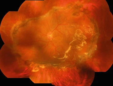
Retinectomy is the surgical removal of a strip of retina to relieve traction when the retina is too short to reattach.
Surgeons use it in severe proliferative vitreoretinopathy and other complex detachments after membrane peeling fails.
Related terms matter, since a relaxing retinotomy cuts the retina without removing it.
Why Retinectomy Is Needed
In PVR, membranes contract the retina and shorten it, even after peeling.
The shortened retina cannot reach the pigment epithelium, so the detachment recurs.
A retinectomy releases that foreshortening and lets the retina lie flat on the posterior eye wall (see proliferative vitreoretinopathy).
Machemer introduced retinotomy techniques in the early 1980s.
Retinectomy Versus Relaxing Retinotomy
A relaxing retinotomy is a circumferential incision of the retina in healthy tissue, and it does not remove tissue.
A retinectomy excises a strip of retina, usually at the edge of the peripheral membranes.
Surgeons use the incision when the shortened area is limited and the removal when the shortened retina covers a wider area.
The extent can reach 360 degrees in severe cases.
Fundus Explorer Pro
Photograph the retinal findings described here with the phone already in your pocket — 22 D optics and built-in illumination in one handheld unit.
From Choroida — the team behind this siteIndications for Retinectomy
- Recurrent PVR detachment with fixed folds
- Giant retinal tears with a rolled posterior edge
- Tractional detachments with anterior loop contraction
- Severe trauma with retinal incarceration
Even so, surgeons try membrane peeling and perfluorocarbon flattening first.
Retinectomy is a salvage step when those fail (see giant retinal tear).
Surgical Technique
The surgeon removes all epiretinal membranes, then identifies the area of shortened retina.
Diathermy marks the retina where the cut will go, and it controls bleeding.
The surgeon then cuts or removes the retina in a controlled arc, using vitrectomy cutter or scissors.
Perfluorocarbon liquid flattens the retina, and the surgeon places laser around the edge of the cut.
A fluid-air exchange follows, with a silicone oil tamponade in most eyes (see silicone oil tamponade complications).
Outcomes of Retinectomy
Anatomic success is possible in a majority of eyes, but visual recovery is limited.
Many patients reach ambulatory vision, and some keep reading vision when the macula stays attached.
Studies of relaxing retinotomy in PVR report reattachment in many eyes with an extended tamponade.
Results vary with the extent of the cut and with the earlier damage.
Complications of Retinectomy
- Bleeding at the cut edge
- Recurrent PVR at the retinectomy margin
- Hypotony and phthisis in severe cases
- Raised pressure or glaucoma with oil
Meticulous diathermy and thorough laser lower the risk of recurrence.
An Example Case
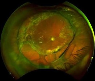
A 52-year-old man has a redetachment with PVR and an anterior fold after two surgeries.
The surgeon performs a 180-degree retinectomy, applies laser, and uses silicone oil.
The retina stays attached for six months, and the surgeon removes the oil with a stable result and 20/200 vision.
Key Points
- Reserve retinectomy for fixed shortening after membrane peeling
- Use diathermy, laser, and long tamponade
- Counsel patients on limited visual gain
Questions Patients Ask
Patients ask whether the surgery will restore reading vision, and the honest answer is that the aim is to save the eye and keep useful vision.
They also ask how long the oil stays, and many keep it for months.
Because outcomes vary, explain that further surgery may follow.
Pitfalls to Avoid
- Cutting before all membranes are removed
- Leaving bleeding vessels at the cut edge
- Skipping laser along the retinectomy margin
- Underestimating the need for long tamponade
Looking Ahead
Researchers test adjuncts such as methotrexate to reduce PVR.
Also, imaging at surgery may guide the extent of retinectomy.
However, these ideas need more evidence, so the basic surgical steps remain the same.
Practical Points
Plan for silicone oil before the case, and have diathermy ready.
Then discuss the possibility of a large retinectomy with the patient in advance.
Also arrange follow-up visits soon after surgery to catch early pressure rises.
Planning the Extent of Retinectomy
Mark the border of healthy retina before cutting.
Also review preoperative imaging, which shows the extent of folds and the position of the macula.
Because the cut should lie posterior to the contracted retina, extend it until the retina lies flat without tension.
Then check the cut edge for residual membranes, since they cause redetachment.
If bleeding occurs, use diathermy at once, and raise the infusion pressure for a moment.
Finally, use enough laser to form a firm adhesion at the cut margin.
Tell the anesthesia team about the expected length of surgery, since a large retinectomy takes time.
Also have silicone oil and long-acting gas available before you start.
For the latest studies, see the PubMed results for retinectomy.


Document what you see
Two smartphone imaging tools built for everyday clinic use — one for the slit lamp, one for the fundus.
From Choroida — the team behind this siteReferences
- Machemer R. Retinotomy. Am J Ophthalmol. 1981;92:768-774.
- Tseng JJ, Barile GR, Schiff WM, et al. Influence of relaxing retinotomy on surgical outcomes in proliferative vitreoretinopathy. Am J Ophthalmol. 2005;140:628-636.
- Ryan SJ, Schachat AP, Wilkinson CP, et al, eds. Retina. 6th ed. Elsevier; 2018.
- American Academy of Ophthalmology. Basic and Clinical Science Course, Section 12: Retina and Vitreous. AAO; current edition.

Writes and reviews the clinical articles on Educate. Every post is checked against peer-reviewed sources before it goes live.