
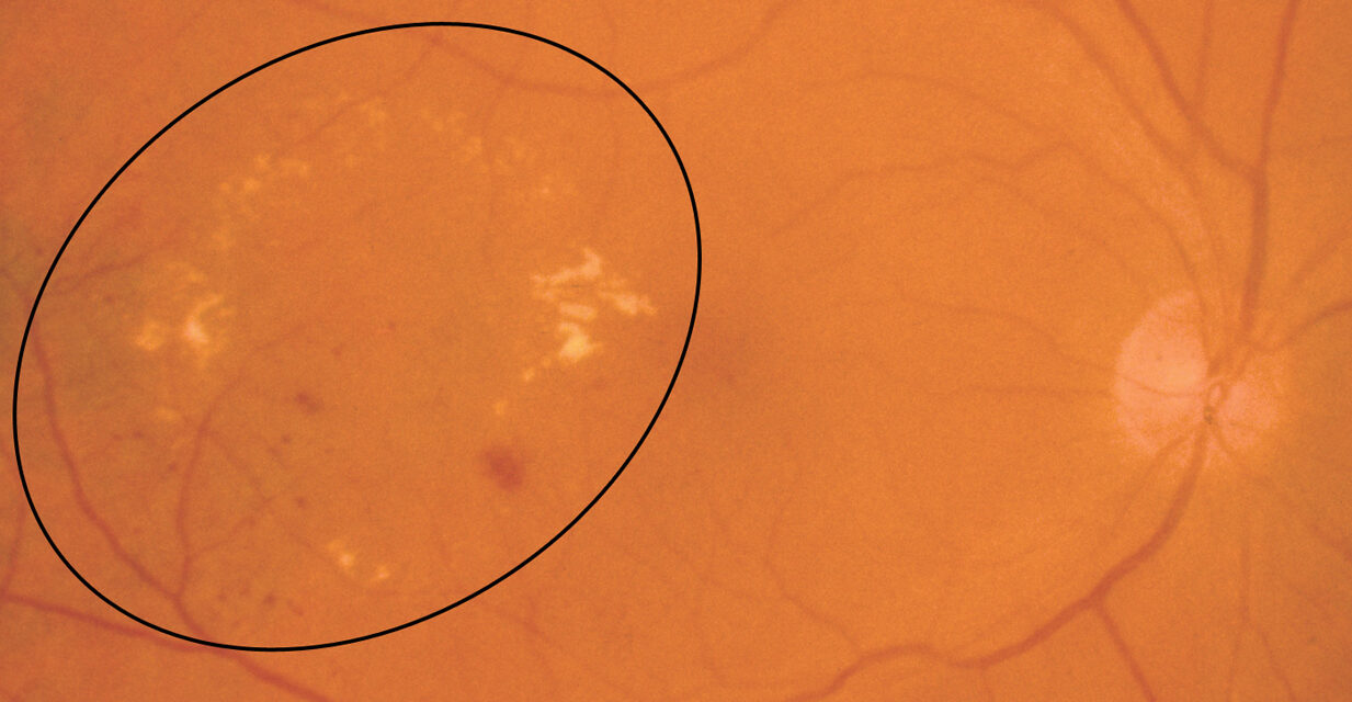
Hard exudates are waxy, yellow-white deposits in the retina with sharp borders.
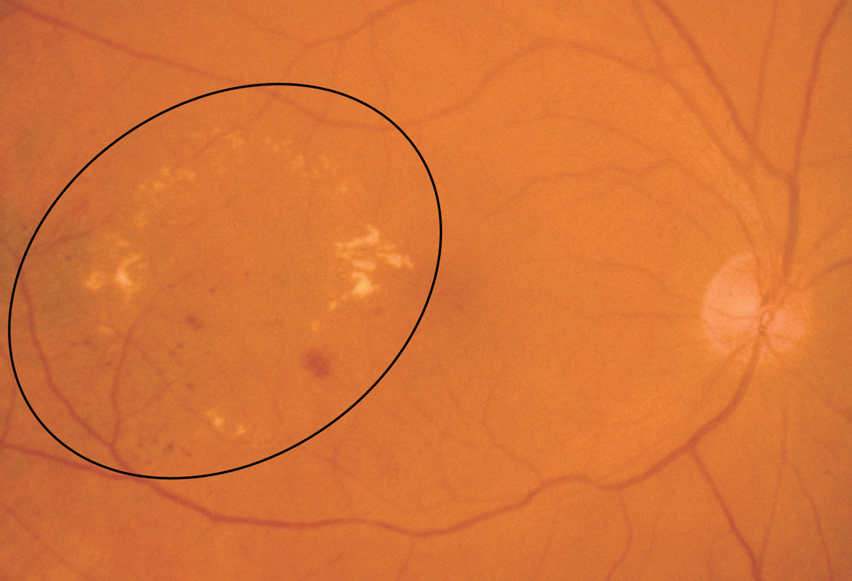
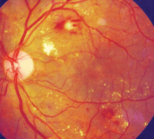
They are not inflammation and not hemorrhage.
They are lipoproteins and lipid-laden macrophages that remain after plasma has leaked from damaged vessels and the watery component has been reabsorbed.
Where they sit and why that matters
The deposits accumulate in the outer plexiform layer, the loosely arranged Henle fiber layer, which is why they take specific shapes.
Around the macula, where the fibers run radially, they form a macular star with a fan of lines pointing at the fovea.
Around a leaking focus such as a microaneurysm or a retinal macroaneurysm, they form a circinate ring, with the leak at the center and the lipid pushed out to the edge of the edema.
What produces them
Any process that makes retinal vessels leak for weeks or months can do it.
- Diabetic retinopathy and diabetic macular edema, by far the commonest cause (see diabetic macular edema)
- Hypertensive retinopathy, where a macular star may appear in accelerated disease (see hypertensive retinopathy)
- Retinal vein occlusion, particularly branch occlusions after the hemorrhage has cleared
- Retinal macroaneurysm
- Coats disease and Coats-like telangiectasia (see Coats-like telangiectasia)
- Neuroretinitis, in which the star appears days after disc swelling begins
- Radiation retinopathy and some inflammatory conditions
Fundus Explorer Pro
Photograph the retinal findings described here with the phone already in your pocket — 22 D optics and built-in illumination in one handheld unit.
From Choroida — the team behind this siteHow to tell them from other yellow lesions
Drusen are deeper, at the level of the RPE, and have softer borders.
Cotton wool spots are fluffy and fade within weeks (see cotton wool spots).
Subretinal lipid after a long-standing exudative detachment sits below the retina and often has a pigmented rim.
OCT settles the question quickly by showing hyperreflective foci in the outer retina.
Management
Hard exudates are a sign, so treatment is aimed at the leaking source.
- Control of glucose, blood pressure and lipids is the foundation
- Focal laser to leaking microaneurysms is still useful for circinate rings away from the fovea
- Anti-VEGF injections are standard when the center of the macula is involved
- Exudates clear slowly, often over many months, and they can leave subretinal fibrosis if foveal deposits have been present for years
Reading the pattern in front of you
The distribution of exudates narrows down the source.
A circinate ring points to a single leaking focus at its center, and fluorescein angiography will usually show the microaneurysm or macroaneurysm.
A diffuse scatter across the posterior pole with microaneurysms and dot hemorrhages is the usual diabetic picture.
A macular star with disc swelling and no diabetes points toward neuroretinitis, and the common infectious cause is Bartonella henselae, with a history of cat contact.
Unilateral dense exudates in a young boy, especially with telangiectatic vessels in the periphery, suggest Coats disease.
A single bilateral macular star with severe hypertension indicates accelerated disease, and these patients need blood pressure management the same day, not an outpatient referral.
Common mistakes
- Treating exudates directly with laser to the deposits themselves. The deposit is the aftermath, and the leaking vessel upstream is the target
- Assuming that clearing of exudates means the macula is healthy. A thick plaque may disappear and leave subfoveal fibrosis and photoreceptor loss
- Missing a hidden source. Extrafoveal exudates with no obvious leaking vessel may reflect a small peripheral telangiectasia or a branch vein occlusion, so scan the periphery
- Ordering lipid-lowering treatment as a replacement for ocular treatment. It helps, particularly with fenofibrate in diabetic retinopathy, but it is an addition
Follow-up and documentation
Record the position of the exudates in relation to the fovea, because a ring that creeps toward the center changes the urgency of treatment. A color photograph and an OCT map at the first visit give a baseline that can be compared after treatment. Review four to twelve weeks after any intervention, since exudates clear slowly and the retinal thickness map improves long before the yellow deposits fade.
Patients should understand that the deposits themselves are not the target, that clearing takes months, and that a reduction in the swelling is the real marker of success. If exudates persist at the fovea after the edema has resolved, tell the patient early that some permanent loss of acuity is likely, so that expectations are realistic.
Prognosis
Peripheral exudates carry no visual cost.
Foveal exudates, particularly a dense plaque that has been there for a long time, tend to limit recovery even after the edema resolves, which is a good reason not to delay treatment.
Typical resolution takes between six and twelve months after the leak is controlled, and patients should be told this at the start so that slow clearing is not mistaken for treatment failure.


Document what you see
Two smartphone imaging tools built for everyday clinic use — one for the slit lamp, one for the fundus.
From Choroida — the team behind this siteReferences
- Bresnick GH. Diabetic maculopathy: a critical review highlighting diffuse macular edema. Ophthalmology. 1983;90:1301-1317.
- Cusick M, Chew EY, Chan CC, Kruth HS, Murphy RP, Ferris FL 3rd. Histopathology and regression of retinal hard exudates in diabetic retinopathy after reduction of elevated serum lipid levels. Ophthalmology. 2003;110:2126-2133.
- Early Treatment Diabetic Retinopathy Study Research Group. Photocoagulation for diabetic macular edema. ETDRS report number 1. Arch Ophthalmol. 1985;103:1796-1806.
- Ryan SJ, ed. Retina. 6th ed. London: Elsevier; 2018.
