
CASE REPORT
A 65-year-old male presented with a two-month history of gradually worsening bilateral blurred vision, associated with a sensation of “thickness” in both eyes and intermittent headaches.


He also reported persistent fatigue and occasional epistaxis. His past medical history revealed controlled hypertension with an angiotensin-converting enzyme inhibitor, with no history of diabetes mellitus or cardiovascular disease.
The patient, a retired school teacher, leads a sedentary lifestyle with no history of tobacco or alcohol use. On examination, he displayed alertness but significant fatigue, and ophthalmologic findings included bilateral dilated retinal veins, flame-shaped hemorrhages, and cotton-wool spots.
Laboratory results indicated mild normocytic anemia, thrombocytopenia, and an elevated monoclonal immunoglobulin M (IgM) spike with increased serum viscosity.
Imaging studies were unremarkable for intracranial abnormalities. The diagnosis of Waldenstrom Macroglobulinemia with Hyperviscosity-Related Retinopathy was established.
DISEASE
Waldenstrom macroglobulinemia (WM) is a malignant lymphoplasmo-proliferative disorder that overproduces IgM, a monoclonal pentameric immunoglobulin.
In patients with WM, the bone marrow or the lymph nodes are occupied by pleomorphic B lymphocytes in various maturation stages ranging from small lymphocytes, and lymphoplasmacytoid cells with basophilic cytoplasm to plasma cells.
Mast cells also increase significantly in the bone marrow of these patients while overexpressing CD40 Ligand, an inducer of B-cell proliferation.
Patients present with cytopenias due to bone marrow infiltration, lytic bone disease, hepatomegaly, splenomegaly, lymphoplasmacytic infiltration of the pulmonary parenchyma, or Bing-Neel syndrome.
Bing-Neel syndrome is a rare complication due to lymphoplasmacytoid infiltration and IgM deposits in the CNS. Perivascular malignant infiltration occurs as a result of long-standing hyperviscosity and secondary CNS vascular hyperpermeability.
Symptoms include vertigo, headache, ataxia, diplopia, nystagmus, and coma. Ocular manifestations of the disease include infiltration of the conjunctivae, malignant vitritis, and hyperviscosity-related retinopathy.
Waldenstrom macroglobulinemia (WM) is primarily considered a sporadic disease, but some studies have suggested a possibility of a single genetic defect.
Deletions in 6q21-22.1 were found in the majority of patients with WM, and B-cell disorders were also found in first-degree relatives of these patients.
Serum hyperviscosity leading to vascular disturbances plays a major role in the microvascular changes of the retina causing vascular dilation.
Physical examination
The physical exam is consistent with hyperviscosity syndrome, presenting with symptoms of neurologic dysfunction such as headache and fatigue, bleeding diathesis such as epistaxis, and visual abnormalities associated with retinopathy.
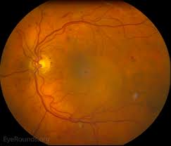
Ocular signs of hyperviscosity manifest as a hyperviscosity-related retinopathy. In the early stages of Waldenstrom macroglobulinemia (WM), it is postulated that small retinal hemorrhages are seen in the far periphery that can be observed on indirect ophthalmoscopy with scleral depression.
Later, the dilated fundoscopic examination also revealed vascular tortuosity and sausage-like dilation of retinal veins, as well as more centrally located retinal hemorrhages, macular edema, and optic disc edema.
The hemorrhages observed are microaneurysms, dot-blot, and flame hemorrhages. It is important to note that retinal hemorrhages can occur without affecting visual acuity if the macula is spared.
Optical coherence tomography (OCT) is often used to monitor macular edema, which can progress to neurosensory serous macular detachments.
Bilateral central retinal vein occlusions, as a result of hyperviscosity syndrome, have also been reported. Fluorescein angiography demonstrates increased retinal circulation time and areas of capillary non-perfusion and microaneurysms.
MANAGEMENT
Initial treatment of Waldenstrom macroglobulinemia (WM) involves managing the acute symptoms due to hyperviscosity syndrome since more than 80% of IgM is intravascular.

Plasmapheresis is used to decrease IgM in the vasculature and is shown to be as effective as reducing serum IgM by 35%-48%. This treatment also helps with reversing retinal findings like vascular engorgement.
Once serum IgM levels are controlled, the next step is to prevent another spike and maintain the levels with chemotherapy.
Cyclophosphamide/doxorubicin/vincristine/prednisone/rituximab (CHOP-R) is a stem cell-sparing regimen, and rituximab (a recent addition to the CHOP regimen) may be added without an increase in toxicity.
Other studies favor the use of DRC (dexamethasone, rituximab, and cyclophosphamide) or BR (bendamustine and rituximab) over CHOP-R as this regimen may be more tolerable and is associated with greater periods of halting disease progression.
Red blood cell transfusions are not recommended due to the risk of increasing serum viscosity from the interaction of host IgM with donor RBCs.
Complications
Life-threatening complications include gastrointestinal bleeding, cerebral hemorrhage, and high-output cardiac failure.
Bilateral central vein occlusions due to compression and engorgement of the retinal venous system and severe macular edema leading to serous macular detachments can lead to permanent vision loss if left untreated.
Prognosis
The median survival of patients with Waldenstrom macroglobulinemia is around five years with up to 40% surviving ten years or more.

In the majority of patients, the cause of death is advanced age-related comorbidities rather than WM itself. The presence of increased immunoblasts/pleomorphic cells, deletion of 6q, and absence of MYD88 L265P mutation are all associated with poor prognosis.
WM can also transform to diffuse large B-cell lymphoma which has an adverse outcome.
Would you have interest in taking retinal images with your smartphone?
Fundus photography is superior to fundus analysis as it enables intraocular pathologies to be photo-captured and encrypted information to be shared with colleagues and patients.
Recent technologies allow smartphone-based attachments and integrated lens adaptors to transform the smartphone into a portable fundus camera and Retinal imaging by smartphone.
RETINAL IMAGING BY YOUR SMARTPHONE
REFERENCES
- Vijay A, Gertz M. Waldenström macroglobulinemia. Blood. 2007;109.12: 5096-5103.
- Hunyor, Alex P. “hyperviscosity-retinopathy.” Retina Image Bank, 2013, imagebank.asrs.org/file/3080/hyperviscosity-retinopathy.
- Menke MN, Feke GT, McMeel JW, Branagan A, Hunter Z, Treon SP. Hyperviscosity-Related Retinopathy in Waldenström Macroglobulinemia. Archives of Ophthalmology. 2006;124(11):1601–1606. doi:10.1001/archopht.124.11.1601
- Cancer.org. “Risk Factors For Waldenstrom Macroglobulinemia.” 2018, cancer.org/cancer/waldenstrom-macroglobulinemia/causes-risks-prevention/risk-factors.
- Stone MJ, Pascual V. Pathophysiology of Waldenström’s macroglobulinemia. Haematologica. 2010;95(3):359-364. doi:10.3324/haematol.2009.017251
- Gertz M. Acute hyperviscosity: Syndromes and management. Blood. 2018 Sep 27; 132(13): 1379–1385
