
CASE REPORT
A 42-year-old man with no known past medical history presented with 3 weeks of blurry vision in the right eye. He had no other ocular, medical, or surgical history. He rarely sought medical care and was on no medications.
Upon further history taking, he endorsed bitemporal headaches over the past 2 weeks and occasional warmth in his face over the same time period.
Ophthalmic examination revealed visual acuity of count fingers in the right eye and 20/20 in the left eye. His intraocular pressure was normal (19 mmHg) in both eyes. His pupils reacted normally without an afferent pupillary defect.
He had normal confrontation visual fields, extraocular motility, and ocular alignment in both eyes. A slit lamp examination revealed normal anterior structures in both eyes.
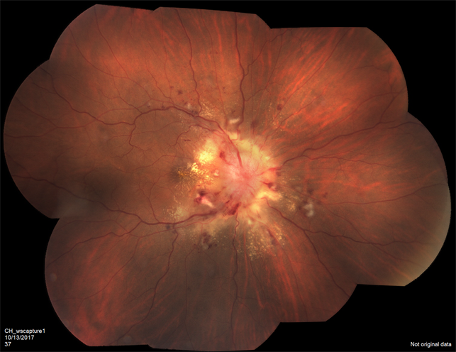
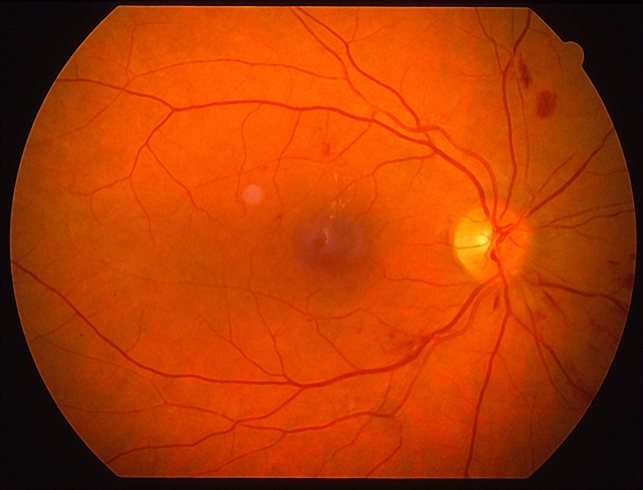
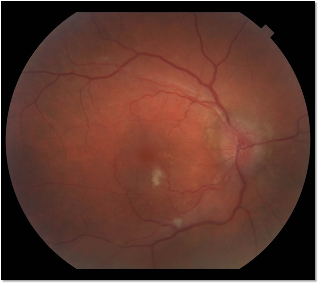
A retinal exam on the right revealed normal optic disc, arteriolar attenuation, diffuse macular and peripapillary cotton wool spots, intraretinal hemorrhages throughout the macula and retinal periphery, intraretinal exudation and macular star formation, and serous retinal detachment of the macula seen clinically and with optical coherence tomography (OCT).
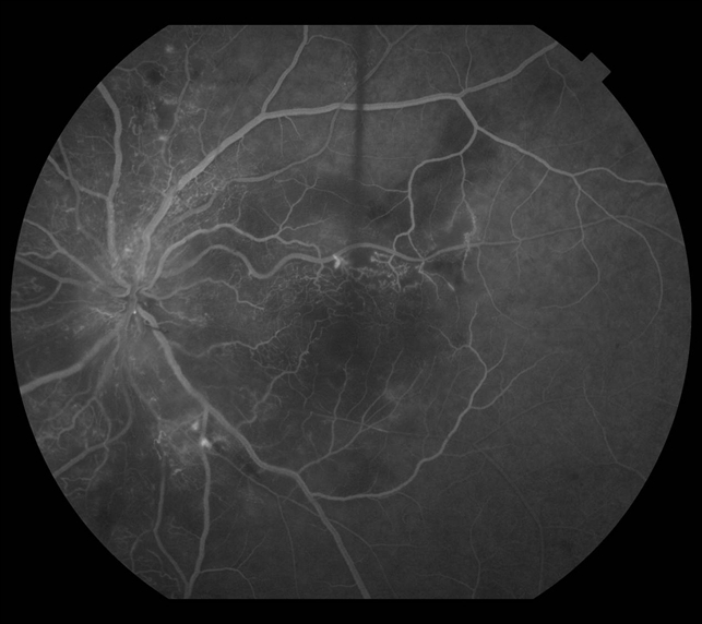
Examination of the left eye revealed similar retinal findings with a lack of exudation and less significant serous retinal detachment also seen on OCT. Fluorescein angiography was performed and demonstrated early hyperfluorescent spots in both eyes with leakage in the late phases.
The patient’s blood pressure was checked and found to be 256/160 mmHg. He was diagnosed with hypertensive retinopathy was confirmed.
Hypertensive Retinopathy DISEASE entity
The arteriosclerotic changes of hypertensive retinopathy are caused by chronically elevated blood pressure. The American College of Cardiology/American Heart Association (ACC/AHA) suggested the following definitions for high blood pressure in 2017.
| Category | Systolic (SBP) | Diastolic (DBP) |
|---|---|---|
| Elevated blood pressure | 120-129 mmHg | < 80 mmHg |
| Stage 1 hypertension | 130-139 mmHg | 80-89 mmHg |
| Stage 2 hypertension | ≥ 140 mmHg | ≥ 90 mmHg |
Hypertensive retinopathy includes two disease processes. The acute effects of systemic arterial hypertension are a result of vasospasm to autoregulate perfusion. The chronic effects of hypertension are caused by arteriosclerosis and predispose patients to visual loss from complications of vascular occlusions or macroaneurysms.
The arteriosclerotic changes of hypertensive retinopathy are caused by chronically elevated blood pressure, defined as SBP greater than 140 mmHg and DBP greater than 90 mmHg. Primary hypertension is usually essential and not secondary to another disease process.
Essential hypertension is a polygenic disease with multiple modifiable environmental factors contributing to the disease. However, secondary hypertension can develop in the setting of pheochromocytoma, primary hyperaldosteronism, Cushing’s syndrome, renal parenchymal disease, renal vascular disease, coarctation of the aorta, obstructive sleep apnea, hyperparathyroidism, and hyperthyroidism.
Many young patients with secondary hypertension may actually present to an ophthalmologist with bilateral vision loss due to serous macular detachment, bilateral optic disc edema, and exudative retinal detachment. In addition, genetic factors have been found to be associated with a higher risk of hypertensive retinopathy.
Fundus Explorer Pro
Photograph the retinal findings described here with the phone already in your pocket — 22 D optics and built-in illumination in one handheld unit.
From Choroida — the team behind this siteMANAGEMENT of Hypertensive Retinopathy
The treatment for hypertensive retinopathy is primarily focused on reducing blood pressure. It is important to work together with the patient’s primary care doctor to ensure timely evaluation and management to reduce ocular and systemic damage.
A thorough history is vital for the proper and timely treatment of hypertensive retinopathy to prevent vision-threatening complications.
General treatment
The treatment for moderate to severe hypertensive retinopathy is to reduce the mean arterial pressure by 10-15% in the first hour. Of note, blood pressure should be controlled by no more than 25% compared to baseline by the end of the first day of treatment to prevent further ischemic damage to target end organs.
Initial treatment often requires parenteral antihypertensive agents and then transitioned to oral agents. Goal-oriented hypertension treatment aims to lower systolic blood pressure to < 130 mmHg and diastolic pressure to < 80 mm Hg over the next 2-3 months.
Medical therapy
Drugs that are commonly used in the outpatient setting to reduce blood pressure include angiotensin-converting enzyme inhibitors, calcium channel blockers, and diuretics. Other less commonly used medications include α-adrenergic blockers, direct vasodilators, and central α2-adrenergic agonists.
The patient should be followed by his primary physician closely for the management of hypertension. If the patient is in a hypertensive crisis, he should be referred to an emergency department for acute management of blood pressure.
Studies have explored intravitreal antibody treatment against vascular endothelial growth factor (bevacizumab) for acute hypertensive retinopathy and showed a reduction in macular edema and retinal hemorrhage.
Moreover, another study showed prompt recovery of malignant hypertensive retinopathy in patients after administering bevacizumab. However, the use of these agents has not yet been widespread or accepted.


Document what you see
Two smartphone imaging tools built for everyday clinic use — one for the slit lamp, one for the fundus.
From Choroida — the team behind this siteREFERENCES
- Tsukikawa, M., & Stacey, A. W. (2020). A Review of Hypertensive Retinopathy and Chorioretinopathy. Clinical optometry, 12, 67–73. https://doi.org/10.2147/OPTO.S183492
- ALEX MELAMUD, PETER K. KAISER, Chapter 28 – Hypertensive Retinopathy; Editor(s): David Huang, Peter K. Kaiser, Careen Y. Lowder, Elias I. Traboulsi, Retinal Imaging, Mosby,2006, Pages 283-288, ISBN 9780323023467
- 2017ACC/AHA/AAPA/ABC/ACPM/AGS/APhA/ASH/ASPC/NMA/PCNA Guideline for the Prevention, Detection, Evaluation, and Management of High Blood Pressure in Adults: A Report of the American College of Cardiology/American Heart Association Task Force on Clinical Practice Guidelines.
- Lang, G.K. Ophthalmology: A Pocket Textbook Atlas (Thieme, Stuttgart, 2007).
- AAO. in Basic and Clinical Sciences Course (Lifelong Education for the Ophthalmologist, San Fransisco, CA, 2006).
- Harjasouliha A, Raiji V, Gonzalez J, Review of hypertensive retinopathy. Dis Mon. 2017 Mar;63(3):63-69.
Test yourself
A few questions straight from this article.
-
What causes the acute retinal effects of systemic arterial hypertension?
Hypertensive retinopathy has two processes: acute changes from vasospasm that autoregulates perfusion, and chronic changes from arteriosclerosis. -
The chronic arteriosclerotic changes of hypertensive retinopathy predispose the eye to which complications?
Chronic hypertension produces arteriosclerosis, which predisposes to visual loss from vascular occlusions or macroaneurysms. -
Under the 2017 ACC/AHA definitions cited for hypertensive retinopathy, what defines stage 2 hypertension?
The ACC/AHA table in the article sets stage 2 hypertension at systolic 140 mmHg or above and diastolic 90 mmHg or above. -
Which macular finding was seen in the right eye of this hypertensive retinopathy case?
The right eye showed cotton wool spots, intraretinal haemorrhages, intraretinal exudation with macular star formation, and a serous macular detachment on OCT. -
What did fluorescein angiography show in this patient with hypertensive retinopathy?
Angiography demonstrated early hyperfluorescent spots in both eyes that leaked in the late phases. -
Which condition is listed as a cause of secondary hypertension in this article?
Secondary hypertension may arise from pheochromocytoma, primary hyperaldosteronism, Cushing's syndrome, renal disease, aortic coarctation, sleep apnoea and thyroid or parathyroid disease. -
How may young patients with secondary hypertension first present to an ophthalmologist?
Many young patients with secondary hypertension present with bilateral vision loss from serous macular detachment, bilateral optic disc oedema and exudative retinal detachment. -
In moderate to severe hypertensive retinopathy, how much should mean arterial pressure fall in the first hour?
Mean arterial pressure should drop 10-15% in the first hour, and by no more than 25% below baseline by the end of the first day to avoid end-organ ischaemia. -
What is the stated blood pressure goal over the two to three months after treating hypertensive retinopathy?
Goal-oriented treatment aims for systolic pressure under 130 mmHg and diastolic under 80 mmHg over the following 2-3 months. -
What have studies of intravitreal bevacizumab in acute hypertensive retinopathy shown?
Bevacizumab studies reported reduced macular oedema and retinal haemorrhage and prompt recovery in malignant hypertensive retinopathy, but such use is not yet widespread or accepted.
