
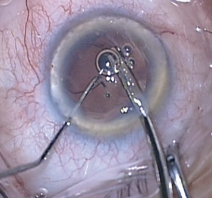
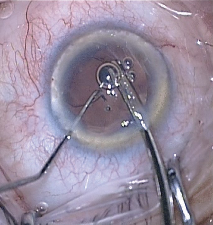
Zonular dehiscence during cataract surgery, whether identified preoperatively or discovered intraoperatively, requires the surgeon to adapt technique in real time to safely remove the lens and achieve stable intraocular lens fixation despite compromised support from the fibers that would normally hold the capsular bag securely centered.
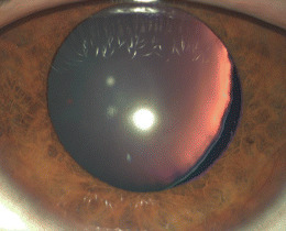
Causes of Zonular Weakness
- Pseudoexfoliation syndrome, one of the most common causes of clinically significant zonular weakness encountered in cataract surgery, related to progressive degeneration of the zonular fibers themselves
- Prior trauma, which can cause focal or more extensive zonular disruption
- Connective tissue disorders, including Marfan syndrome and homocystinuria, associated with generalized zonular weakness and ectopia lentis
- High myopia, associated with a degree of zonular laxity in some patients
- Prior intraocular surgery or chronic inflammation
- Age-related zonular changes, occurring to some degree in the general older population but more pronounced in the conditions above
Preoperative Recognition
- Phacodonesis (visible trembling of the lens with eye movement) and iridodonesis (trembling of the iris, reflecting the same underlying lack of firm zonular support) are important preoperative signs of zonular compromise
- Visible pseudoexfoliation material on the lens capsule or pupillary margin
- A history of trauma or a known connective tissue disorder
- Asymmetric anterior chamber depth or a decentered lens, suggesting focal zonular weakness
Recognizing these signs preoperatively allows for surgical planning, patient counseling about increased complexity and risk, and preparation of specialized equipment that might be needed.
Fundus Explorer Pro
Photograph the retinal findings described here with the phone already in your pocket — 22 D optics and built-in illumination in one handheld unit.
From Choroida — the team behind this siteIntraoperative Recognition
Even without preoperative signs, zonular dehiscence can be discovered intraoperatively, indicated by unexpected lens or capsular bag instability, a wrinkling or striae pattern in the anterior capsule during capsulorhexis, or vitreous presenting unexpectedly at the equator of the lens.
Prompt recognition of intraoperative zonular compromise, rather than proceeding with standard technique as if the support were normal, is essential to avoiding more significant complications, including posterior capsular rupture and lens fragment dislocation into the vitreous.
Intraoperative Management Strategies
Capsular Support Devices
- Capsular tension rings, placed within the capsular bag, help distribute residual zonular support more evenly around the bag’s circumference and provide mechanical stability during phacoemulsification and lens implantation, particularly valuable for mild to moderate zonular weakness (see capsular tension ring indications)
- Capsular hooks or iris hooks can be used to mechanically stabilize the capsular bag margin directly during surgery in eyes with more focal or severe zonular compromise
Modified Surgical Technique
- Gentle, low-flow phacoemulsification technique, minimizing stress on the already compromised zonular support
- A smaller, more controlled capsulorhexis, since a capsulorhexis that is too large in a zonulopathic eye increases the risk of further zonular stress during lens removal
Managing More Severe Dehiscence
For zonular dehiscence too extensive to adequately stabilize with a standard capsular tension ring, options include a modified capsular tension ring with fixation hooks or eyelets that can be sutured to the sclera, or, for the most severe cases, planned conversion to an alternative intraocular lens fixation strategy, including sutured posterior chamber lens fixation or an anterior chamber lens, when capsular bag support proves inadequate for standard in-the-bag lens placement.
Postoperative Considerations
Eyes with significant zonular compromise require closer postoperative monitoring for lens or capsular bag decentration or subluxation over time, since even a lens successfully placed at the time of surgery can gradually decenter if the underlying zonular support continues to weaken.
Prognosis
With appropriate preoperative or intraoperative recognition and the use of capsular support devices and modified technique, most eyes with zonular dehiscence achieve stable, well-centered intraocular lens placement and good visual outcomes.
The greatest risk lies in unrecognized or inadequately managed zonular compromise, which can lead to posterior capsular rupture, lens fragment loss into the vitreous, and a more complex, higher-risk surgical course than would be needed with earlier recognition and appropriate technique adaptation.


Document what you see
Two smartphone imaging tools built for everyday clinic use — one for the slit lamp, one for the fundus.
From Choroida — the team behind this siteReferences
- Ahmed IIK, Chen SH, Kranemann C, Wong DT. Surgical repositioning of dislocated capsular tension rings. Ophthalmology. 2005;112:1725-1733.
- Cionni RJ, Osher RH, Marques DM, Marques FF, Snyder ME, Shapiro S. Modified capsular tension ring for patients with congenital loss of zonular support. J Cataract Refract Surg. 2003;29:1668-1673.
- Hasanee K, Ahmed II. Capsular tension rings: update on endocapsular support devices. Ophthalmol Clin North Am. 2006;19:507-518.
- Jacob S, Agarwal A, Agarwal A, et al. Efficacy of a capsular tension ring for phacoemulsification in eyes with zonular dialysis. J Cataract Refract Surg. 2003;29:315-321.
Test yourself
A few questions straight from this article.
-
Which condition is among the most common causes of clinically significant zonular weakness at cataract surgery?
Pseudoexfoliation syndrome is one of the commonest causes of clinically significant zonular weakness met in cataract surgery, through progressive degeneration of the zonular fibres themselves. -
Which connective tissue disorders cause generalised zonular weakness with ectopia lentis?
Marfan syndrome and homocystinuria are the connective tissue disorders named as causing generalised zonular weakness and ectopia lentis. -
Iridodonesis found before cataract surgery reflects which underlying problem?
Iridodonesis, trembling of the iris, reflects the same lack of firm zonular support that produces phacodonesis, and both are important preoperative signs of zonular compromise. -
During capsulorhexis, which anterior capsule appearance suggests unrecognised zonular dehiscence?
Zonular dehiscence may be discovered intraoperatively through unexpected bag instability, a wrinkling or striae pattern in the anterior capsule during capsulorhexis, or vitreous presenting at the lens equator. -
Why must zonular compromise be recognised promptly during cataract surgery?
Proceeding with standard technique as if support were normal risks more significant complications, including posterior capsular rupture and dislocation of lens fragments into the vitreous. -
What does a capsular tension ring achieve in an eye with mild to moderate zonular weakness?
Placed inside the capsular bag, the ring distributes residual zonular support more evenly around the bag circumference and gives mechanical stability during phacoemulsification and lens implantation. -
How should the capsulorhexis be sized in an eye with known zonular weakness?
A capsulorhexis that is too large in a zonulopathic eye raises the risk of further zonular stress during lens removal, so a smaller, more controlled opening is made. -
Which options suit zonular dehiscence too extensive for a standard capsular tension ring?
Options include a modified capsular tension ring with hooks or eyelets sutured to the sclera, or in the most severe cases a sutured posterior chamber lens or an anterior chamber lens. -
Why do eyes with significant zonular compromise need closer postoperative monitoring?
Even a lens placed successfully at surgery can gradually decentre or subluxate if the underlying zonular support continues to weaken over time. -
What outcome is expected when zonular dehiscence is recognised and managed appropriately?
With recognition and the use of capsular support devices and modified technique, most eyes with zonular dehiscence achieve stable, well-centred lens placement and good visual outcomes.
