
Chorioretinal biopsy obtains tissue directly from the retina and choroid for histopathologic or molecular analysis, reserved for a genuinely small subset of cases where less invasive diagnostic methods, including imaging, blood testing, and vitreous or aqueous sampling, have failed to establish a diagnosis for an atypical or progressive intraocular process.
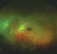
Because the retina and choroid are delicate, functionally irreplaceable tissue, biopsy here is approached with a level of caution rarely needed elsewhere in ophthalmology, and it is pursued only when the diagnostic uncertainty is significant enough, and the consequences of remaining undiagnosed serious enough, to justify the inherent risk to vision the procedure carries.
Understanding when this genuinely last-resort diagnostic tool is appropriate, and how it differs from far more commonly performed vitreous biopsy, clarifies why it remains such an uncommon procedure even at major referral centers.
Most vitreoretinal surgeons will perform this specific procedure only rarely across an entire career, reflecting both how narrow its indications are and how seriously the risk-benefit balance is weighed before proceeding.
Why This Is a Last-Resort Procedure
Unlike a vitreous or aqueous tap, which samples fluid without disturbing functional retinal tissue, a true chorioretinal biopsy removes a small piece of the retina and choroid themselves, tissue with no capacity to regenerate and directly responsible for the vision in the biopsied area.
This makes chorioretinal biopsy fundamentally different in risk profile from the vitreous biopsy techniques used far more commonly to diagnose infectious endophthalmitis or intraocular lymphoma, discussed in their own dedicated articles on this site, which sample fluid rather than functional tissue directly.
Given this risk, the procedure is reserved for cases where the diagnostic uncertainty carries serious consequences, most often distinguishing between a malignancy, a specific infection, or a rare inflammatory condition when this distinction cannot be reached through any less invasive means.
Indications
- Suspected intraocular lymphoma or other malignancy where vitreous biopsy has been non-diagnostic or is not technically feasible, and the diagnostic uncertainty carries major treatment implications
- An atypical, progressive chorioretinal infiltrate or lesion of unclear cause despite thorough systemic and vitreous-based workup
- Suspected rare infectious causes not identifiable through less invasive sampling, where tissue architecture and organism location within the tissue itself may aid diagnosis in a way fluid sampling alone cannot
- Atypical presentations of white dot syndromes or other inflammatory chorioretinal conditions where malignancy cannot otherwise be confidently excluded
Fundus Explorer Pro
Photograph the retinal findings described here with the phone already in your pocket — 22 D optics and built-in illumination in one handheld unit.
From Choroida — the team behind this siteTechnique
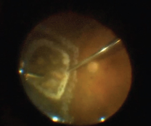
The procedure is typically performed as part of a pars plana vitrectomy, discussed in its own dedicated article on this site, with the biopsy itself obtained from a carefully selected area, generally chosen at the edge of an active lesion where diagnostic yield is expected to be highest.
Endolaser or cryotherapy is typically applied around the biopsy site both before and after tissue removal to minimize bleeding and help seal the retina, given the inherent risk of retinal detachment at a site where full-thickness tissue has been deliberately removed.
The obtained tissue undergoes histopathologic examination and, increasingly, molecular and genetic analysis, which can provide diagnostic information beyond what traditional light microscopy alone would reveal, particularly relevant for lymphoma subtyping and certain infectious diagnoses.
Site Selection
Choosing the biopsy location carefully, generally favoring peripheral over central retina when the disease process allows, balances diagnostic yield against the visual consequences of removing tissue from a given area.
When the disease process is confined to or most active in the macula, a genuine tension exists between obtaining diagnostically useful tissue and preserving central vision, and this decision requires careful, individualized discussion with the patient about the trade-offs involved.
Risks
Retinal detachment at or near the biopsy site is the most significant procedure-specific risk, reflecting the deliberate full-thickness tissue removal the technique requires.
Vitreous hemorrhage, and permanent visual field loss corresponding to the biopsied area, are additional expected consequences of the procedure that need to be clearly discussed with the patient beforehand.
Despite careful technique, the biopsy can still be non-diagnostic, particularly if the sampled area does not adequately represent the underlying disease process, a real possibility given how small the sampled tissue typically is relative to the entire chorioretinal disease process being investigated.
This possibility of a non-diagnostic result, despite the real risk incurred to obtain it, is a genuinely difficult part of the informed consent conversation and deserves honest, explicit discussion before the procedure rather than being glossed over.


Document what you see
Two smartphone imaging tools built for everyday clinic use — one for the slit lamp, one for the fundus.
From Choroida — the team behind this siteReferences
- Manjandavida FP, Shields CL. The role of chorioretinal biopsy in the diagnosis of intraocular tumors and inflammatory conditions. Indian Journal of Ophthalmology.
- American Academy of Ophthalmology. Basic and Clinical Science Course, Section 12: Retina and Vitreous.
- Augsburger JJ, Greatrex KV. Fine needle aspiration biopsy of solid intraocular tumors: indications, instrumentation and techniques. Ophthalmic Surgery.
Test yourself
A few questions straight from this article.
-
When is chorioretinal biopsy considered in an atypical intraocular process?
Chorioretinal biopsy is reserved for the small subset of cases where less invasive methods, including imaging, blood testing and vitreous or aqueous sampling, have not established a diagnosis. -
What makes chorioretinal biopsy riskier than a vitreous or aqueous tap?
A tap samples fluid without disturbing functional tissue, whereas chorioretinal biopsy removes retina and choroid, tissue with no capacity to regenerate and directly responsible for vision in that area. -
Which scenario is an accepted indication for chorioretinal biopsy?
Suspected lymphoma or other malignancy where vitreous biopsy has been non-diagnostic or is not technically feasible, and the uncertainty carries major treatment implications, is an accepted indication. -
From which part of a chorioretinal lesion is the biopsy specimen generally taken?
The specimen is generally taken from the edge of an active lesion, where diagnostic yield is expected to be highest. -
Why is endolaser or cryotherapy applied around a chorioretinal biopsy site?
Endolaser or cryotherapy is applied around the site both before and after tissue removal to minimise bleeding and help seal the retina, given the detachment risk at a full-thickness defect. -
As part of which operation is chorioretinal biopsy typically performed?
The biopsy is typically obtained as part of a pars plana vitrectomy, with the sample taken from a carefully selected area of the lesion. -
What does molecular and genetic analysis add to chorioretinal biopsy tissue?
Alongside histopathology, molecular and genetic analysis can reveal diagnostic information traditional light microscopy would miss, which is particularly relevant for lymphoma subtyping and certain infections. -
Which biopsy site is generally preferred when the disease process allows a choice?
Site selection generally favours peripheral over central retina when the disease process allows, balancing diagnostic yield against the visual consequences of removing tissue. -
What is the most significant procedure-specific risk of chorioretinal biopsy?
Retinal detachment at or near the biopsy site is the most significant procedure-specific risk, reflecting the deliberate full-thickness tissue removal the technique requires. -
Which outcome must be explicitly discussed during consent for chorioretinal biopsy?
The sampled tissue may not adequately represent the underlying disease, so a non-diagnostic result remains possible despite the real risk incurred, and this deserves honest, explicit discussion beforehand.
