
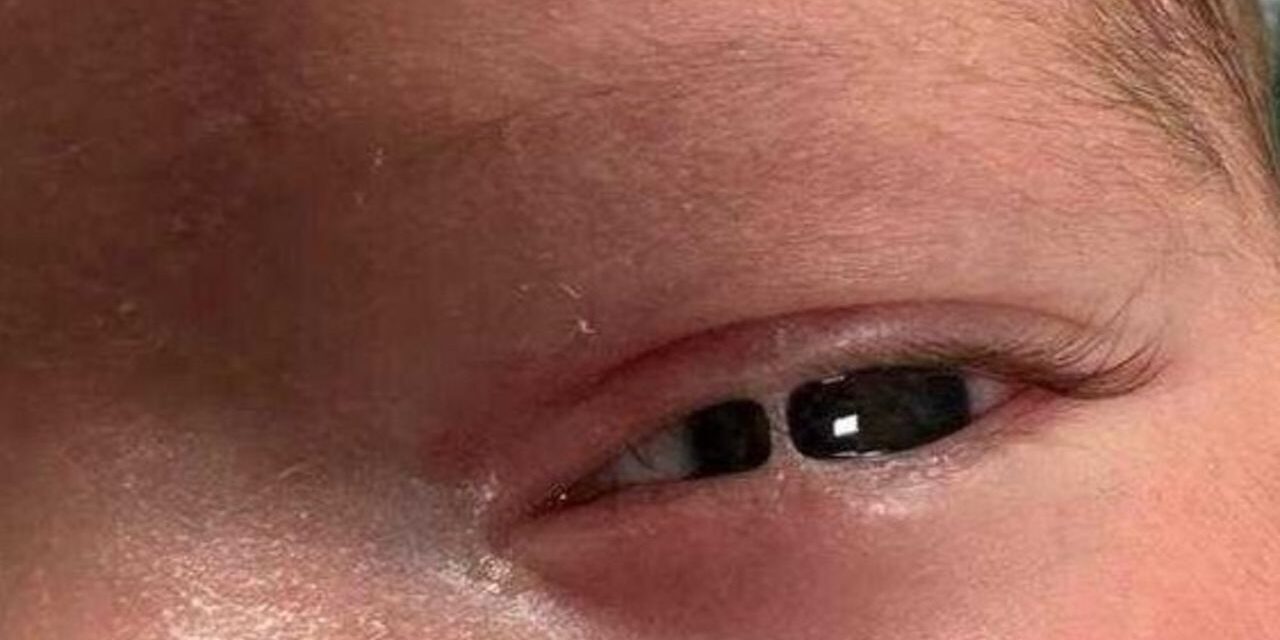
Ankyloblepharon is partial or complete fusion of the upper and lower eyelid margins, distinct from symblepharon, where it is the conjunctiva that adheres abnormally, typically between the palpebral and bulbar surfaces rather than the lid margins themselves.
The congenital form is a distinct entity from the acquired form seen after severe cicatrizing disease, and the two differ enough in cause and management that they are best thought of separately rather than as points on a single spectrum.
Both share the same basic anatomic problem, an abnormal bridge of tissue holding the lids together, but the underlying reason that bridge formed changes everything about how it is approached.
Ankyloblepharon in its congenital, isolated form is genuinely rare, which means most clinicians will encounter it only a handful of times, if ever, across a career.
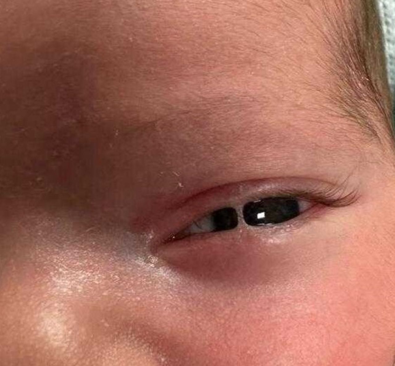
Congenital Ankyloblepharon
Congenital ankyloblepharon filiforme adnatum is a rare condition in which fine, thread-like bands of tissue connect the upper and lower lid margins at birth, most often at the lateral canthus, sometimes limiting how widely the eye can open.
It can occur as an isolated finding or as part of a recognized syndrome, including Hay-Wells syndrome, also known as AEC syndrome, which combines ankyloblepharon with ectodermal dysplasia and cleft lip or palate.
Because a significant minority of cases are syndromic, a newborn with ankyloblepharon warrants a broader examination for associated skin, hair, dental, and craniofacial abnormalities rather than assuming an isolated, incidental finding.
Other described associations include popliteal pterygium syndrome and CHAND syndrome, both rare, which is one more reason a pediatric genetics referral is often appropriate once the finding is confirmed.
Acquired Ankyloblepharon
- Severe cicatrizing conjunctival disease, including ocular cicatricial pemphigoid and Stevens-Johnson syndrome, both discussed in their own dedicated articles on this site, which can progress to lid margin fusion in severe, longstanding cases
- Chemical or thermal burns severe enough to destroy the lid margin epithelium on both lids simultaneously
- Chronic, severe blepharitis or infection left untreated over an extended period
- Prior eyelid surgery or trauma involving both lid margins in the same area
Acquired ankyloblepharon is a marker of how severe and longstanding the underlying disease process has been, since simple inflammation alone rarely progresses this far without significant, prolonged epithelial breakdown on both apposing lid surfaces.
Seeing it should prompt a broader assessment of the rest of the ocular surface for other signs of severe cicatrization, since lid margin fusion rarely occurs as an isolated finding in an otherwise mildly affected eye.
All-fit Slit-Lamp Adapter
Record and share exactly what you see at the slit lamp. One adapter fits any slit lamp or surgical microscope — and any smartphone.
From Choroida — the team behind this siteClinical Presentation
In the congenital form, the fusion is typically noted immediately at birth, sometimes limiting the newborn eye exam and prompting early separation specifically to allow adequate visual assessment and screening for other ocular anomalies.
In the acquired form, the fusion develops gradually over the course of the underlying cicatrizing disease, often alongside other signs of severe ocular surface scarring such as symblepharon and, in advanced cases, a severely dry, keratinized ocular surface.
Functional consequences depend on the extent of fusion: a small filiform band may cause minimal visual impact, while more extensive fusion can meaningfully restrict lid opening and, in an infant, raise real concern for deprivation amblyopia if not addressed promptly.
Management
Congenital filiform bands are typically simple to separate surgically, often in the office or with brief sedation, since the bands themselves are usually thin, avascular, and straightforward to divide without significant bleeding.
Any newborn with ankyloblepharon should also be evaluated for associated syndromic features, since identifying a condition such as Hay-Wells syndrome has implications well beyond the eyelids themselves.
Acquired ankyloblepharon from cicatrizing disease requires treatment of the underlying condition alongside surgical separation, since dividing the fusion without controlling active inflammation invites recurrence.
Reconstructive surgery for extensive acquired fusion is more complex than for the thin congenital bands, sometimes requiring mucous membrane grafting to restore functional lid margins and prevent the fused tissue from simply reforming during healing.
Postoperative care after separating any fusion also matters as much as the procedure itself, since the two raw, apposing surfaces will tend to re-adhere during healing unless actively kept apart with lubrication, a spacer, or a similar mechanical barrier during the critical early period.


Document what you see
Two smartphone imaging tools built for everyday clinic use — one for the slit lamp, one for the fundus.
From Choroida — the team behind this siteReferences
- McNamara EK, Papapavlou G, Vollmer-Snarr HR, et al. Hay-Wells syndrome: a case series and review of the literature. Ophthalmic Plastic and Reconstructive Surgery.
- American Academy of Ophthalmology. Basic and Clinical Science Course, Section 6: Pediatric Ophthalmology and Strabismus.
- Bowling B. Kanski’s Clinical Ophthalmology.
Test yourself
A few questions straight from this article.
-
Ankyloblepharon is defined as fusion of which ocular structures?
Ankyloblepharon is partial or complete fusion of the upper and lower eyelid margins, held together by an abnormal bridge of tissue. -
Which adhesion defines symblepharon rather than ankyloblepharon?
In symblepharon it is the conjunctiva that adheres abnormally, typically between the palpebral and bulbar surfaces, rather than the lid margins fusing to each other. -
Where are the thread-like bands of ankyloblepharon filiforme adnatum most often found?
Congenital ankyloblepharon filiforme adnatum presents at birth with fine thread-like bands joining the lid margins, most often at the lateral canthus. -
Hay-Wells syndrome combines ankyloblepharon with which two features?
Hay-Wells syndrome, also called AEC syndrome, pairs ankyloblepharon with ectodermal dysplasia and cleft lip or palate. -
A newborn found to have ankyloblepharon warrants examination for abnormalities of what?
A significant minority of congenital cases are syndromic, so the newborn needs a broader look for associated skin, hair, dental and craniofacial abnormalities. -
Which disease process leads to acquired ankyloblepharon?
Severe cicatrizing conjunctival disease, including ocular cicatricial pemphigoid and Stevens-Johnson syndrome, can progress to lid margin fusion when severe and longstanding. -
What must accompany surgical separation in acquired ankyloblepharon from cicatrizing disease?
Dividing the fusion without controlling the active inflammation simply invites recurrence, so the underlying disease has to be treated alongside surgery. -
Extensive ankyloblepharon restricting lid opening in an infant raises concern for what?
Fusion extensive enough to meaningfully restrict lid opening in an infant carries real risk of deprivation amblyopia unless it is addressed promptly. -
Why are congenital filiform bands usually straightforward to divide surgically?
The bands are typically thin and avascular, which allows separation in the office or under brief sedation without significant bleeding. -
What is essential after separating fused eyelid margins to stop the fusion reforming?
The two raw apposing surfaces tend to re-adhere while healing, so they must be actively kept apart with lubrication, a spacer or a similar mechanical barrier early on.
