
DISEASE
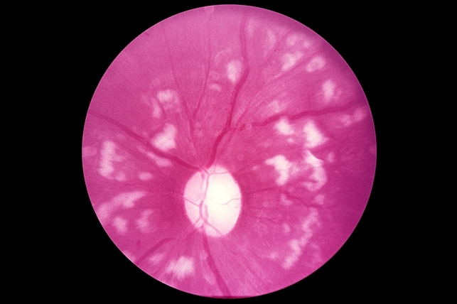
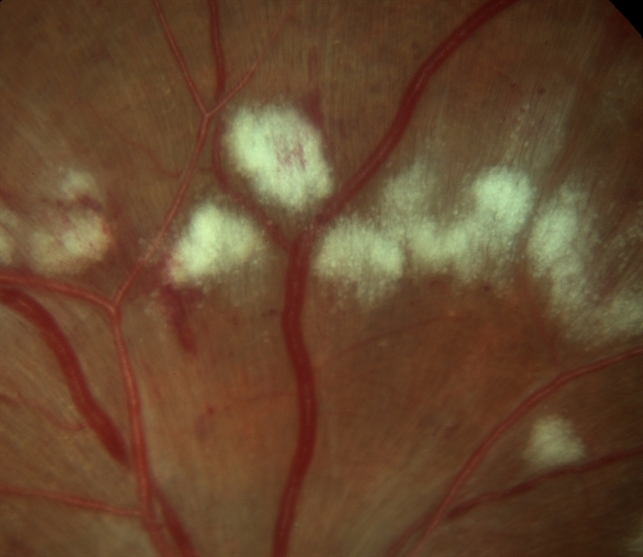
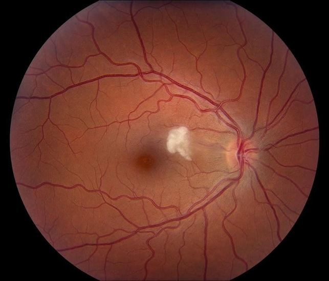
In otherwise healthy patients, the observance of a cotton wool spot (CWS) is not considered normal. A single cotton wool spot in one eye can be the earliest ophthalmoscopic finding in diabetic or hypertensive retinopathy.
In a series of patients who had (CWS) and no known medical history, diastolic blood pressure equal to or greater than 90 mmHg was detected in 50% of patients, and elevated blood sugar was found in 20% of patients.
Pathophysiology
Cotton wool spots are believed to occur secondary to ischemia from retinal arteriole obstruction. It is thought to represent nerve fiber layer infarct and pre-capillary arteriolar occlusion.
However, factors responsible for focal interruption of axoplasmic flow in the retinal nerve fiber layer may result in similar intra-axonal organelle accumulations.
The histological hallmark of cotton wool spots is considered by many authors to be cytoid bodies. They are named cytoid bodies because they look like cells,
However, they are eosinophilic segments of ganglion cell axons that are swollen because of defective axoplasmic flow. Cytoid bodies are usually packed with accumulations of mitochondria and other intracellular material.
Fundus Explorer Pro
Photograph the retinal findings described here with the phone already in your pocket — 22 D optics and built-in illumination in one handheld unit.
From Choroida — the team behind this siteMANAGEMENT
Workup and treatment are directed towards the underlying etiology. Cotton wool spots classically disappear in 6–12 weeks,
However, in diabetic retinopathy, they may persist for longer.
Initial work-up may include vitals (blood pressure, heart rate, etc.) and metabolic studies including HbA1c, CBC, CMP, and HIV.
Directed work-up may include ESR, CRP, EKG, echocardiogram, carotid ultrasound, hypercoagulable labs (protein C, protein S, Leiden, etc), PT/PTT, homocysteine, and more.
REFERENCES
- Adam T. Gerstenblith, Michael P. Rabinowitz. Wills Eye Manual, The: office and emergency room diagnosis and treatment of eye disease, 6th edition. Lippincott Williams & Wilkins, 2012.
- Brown GC, Brown MM, Hiller T, Fischer D, Benson WE, Magargal LE. “Cotton-wool spots.” Retina. 5, no. 4 (Fall-Winter 1985): 206-14.
- Arroyo, Jorge G. “Cotton-Wool Spots May Challenge Diagnosis .” Review of ophthalmology, 2004: 111-114.
- Arroyo JG, Irvine AR. “Retinal distortion and cotton-wool spots associated with epiretinal membrane contraction.” Ophthalmology 102 (1995): 662-8.
- Purnima S. Patel, SriniVas R. Sadda. “Retinal Artery Obstructions.” In Retina (Fifth Edition), by Stephen J. Ryan, 1012-1025. Elsevier Inc., 2012.D. McLeod, D.
- McLeod, E. M. Kohner and A. C. Bird. “The role of axoplasmic transport in the pathogenesis of retinal cotton-wool spots.” Br J Ophthalmol 61 (1977): 177-191.


Document what you see
Two smartphone imaging tools built for everyday clinic use — one for the slit lamp, one for the fundus.
From Choroida — the team behind this siteRelated Reading
Test yourself
A few questions straight from this article.
-
What does a single cotton wool spot in one eye of an otherwise healthy patient suggest?
A cotton wool spot is never a normal finding in a healthy patient, and a single spot in one eye can be the earliest ophthalmoscopic sign of diabetic or hypertensive retinopathy. -
In a series of patients with cotton wool spots and no known medical history, how many had a diastolic blood pressure of 90 mmHg or more?
Half of these patients were found to have a diastolic blood pressure of 90 mmHg or higher, and 20% had elevated blood sugar, which is why vitals and metabolic testing come first. -
What retinal event is a cotton wool spot believed to represent?
Cotton wool spots are thought to arise from ischaemia due to retinal arteriole obstruction, representing a nerve fibre layer infarct with pre-capillary arteriolar occlusion. -
Besides ischaemia, which process can produce the same retinal appearance as a cotton wool spot?
Any factor that focally interrupts axoplasmic flow within the retinal nerve fibre layer can produce similar intra-axonal accumulations of organelles. -
What is regarded as the histological hallmark of a cotton wool spot?
Many authors consider cytoid bodies to be the histological hallmark of a cotton wool spot; they are named for their resemblance to cells. -
What are the cytoid bodies seen within a cotton wool spot?
Despite looking like cells, cytoid bodies are eosinophilic segments of ganglion cell axons swollen by defective axoplasmic flow and packed with mitochondria and other intracellular material. -
Over what period do cotton wool spots classically fade?
Cotton wool spots classically disappear over 6 to 12 weeks, although in diabetic retinopathy they may persist for considerably longer. -
What is the guiding principle of management once cotton wool spots are found?
There is no treatment for the spot itself; the workup and treatment are directed towards the underlying systemic aetiology that produced the ischaemia. -
Which investigations belong to the initial work-up of a patient with cotton wool spots?
The initial work-up covers vitals such as blood pressure and heart rate, together with metabolic studies including HbA1c, CBC, CMP and HIV testing. -
Which test belongs to the directed, second-line work-up for cotton wool spots?
Directed testing may add carotid ultrasound, echocardiography, EKG, ESR and CRP, hypercoagulable studies, PT/PTT and homocysteine once the initial screen points that way.

Writes and reviews the clinical articles on Educate. Every post is checked against peer-reviewed sources before it goes live.


