Minimally invasive glaucoma surgery lowers intraocular pressure through small incisions with a better safety profile than classic filtering surgery.
It is also most often combined with cataract surgery in patients with mild to moderate glaucoma.
The effect is more modest than that of trabeculectomy, but recovery is faster, and complications are fewer (see trabeculectomy).
Why Minimally Invasive Glaucoma Surgery Was Developed
Trabeculectomy and tube shunts lower pressure well, but they carry risks, such as hypotony, infection, and scarring, and need long follow-up (see glaucoma drainage devices).
Drops are effective, but many patients do not use them regularly, and have side effects.
Also, MIGS fills the gap between drops and filtering surgery.
Minimally Invasive Glaucoma Surgery: Trabecular Bypass Devices
- iStent and iStent inject: tiny titanium stents placed into Schlemm canal through the trabecular meshwork, which increase outflow (see the references)
- Hydrus microstent: a flexible scaffold that holds open about three clock hours of the canal
Trials, such as the HORIZON study of Hydrus with cataract surgery, showed greater freedom from drops and lower pressure than cataract surgery alone (see the references).
All-fit Slit-Lamp Adapter
Record and share exactly what you see at the slit lamp. One adapter fits any slit lamp or surgical microscope — and any smartphone.
From Choroida — the team behind this siteProcedures That Remove or Cut the Meshwork
- Goniotomy, using devices such as the Kahook dual blade, which removes a strip of trabecular meshwork
- Gonioscopy-assisted transluminal trabeculotomy: opens the canal along its full length
- Canaloplasty: dilates and tensions the canal
These procedures keep tissue open without implants, and may also give a larger drop in pressure.
Minimally Invasive Glaucoma Surgery: Subconjunctival Devices
The XEN gel stent and the PreserFlo microshunt create a pathway from the anterior chamber to the subconjunctival space, resembling a small filtering bleb (see the references).
They can lower pressure more, but they carry bleb-related risks, and need needling in some cases.
Suprachoroidal Devices
Stents placed in the suprachoroidal space lowered pressure, but one device was withdrawn after long-term loss of corneal endothelial cells, a lesson about safety monitoring.
Who Is a Candidate?
- Mild to moderate open-angle glaucoma
- Patients having cataract surgery who want to reduce drops
- Patients intolerant of drops
- Eyes with open angles, and no need for very low target pressure
Eyes with advanced damage and high pressures, however, may need trabeculectomy or a tube.
Minimally Invasive Glaucoma Surgery: Results and Limits
- Most patients reduce the number of drops and the pressure by about one fifth to one third
- Success varies with the type of device and baseline pressure
- The effect may also wane over time, so some eyes need further treatment
- Long-term data are limited for many devices
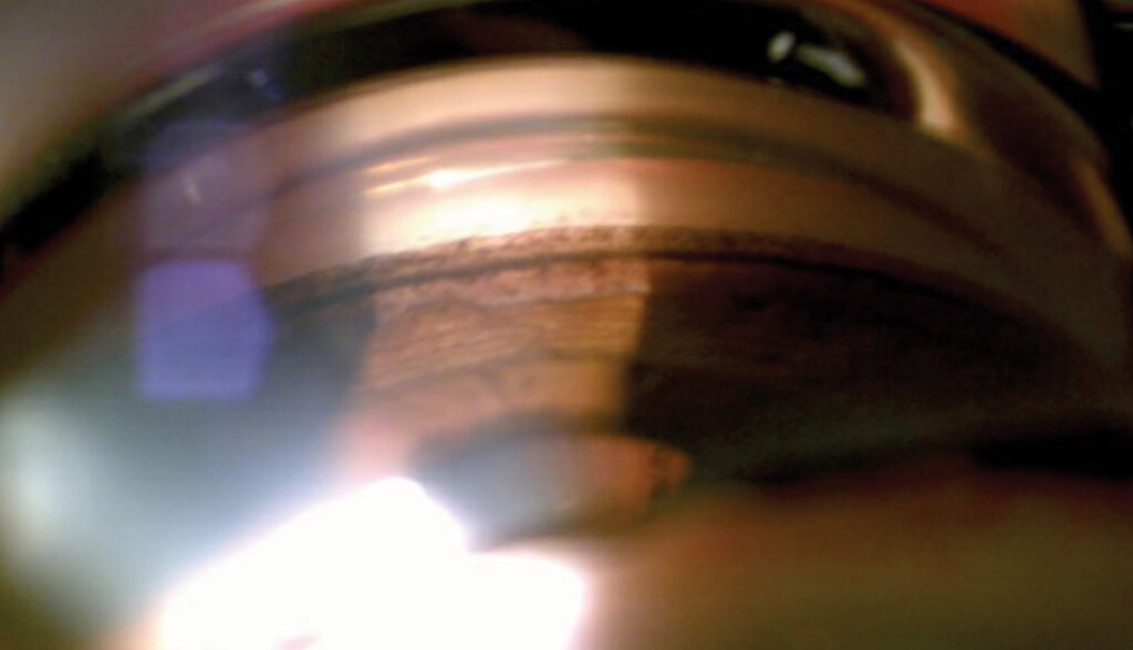
Complications of Minimally Invasive Glaucoma Surgery
- Bleeding in the anterior chamber, often minor
- Malposition of the stent, and obstruction
- Transient pressure spikes and hypotony
- Rarely, corneal endothelial loss or infection
Counselling
Explain that the goal is to lower pressure and drop burden, not to cure glaucoma.
Patients still need regular visual field and pressure checks (see primary open-angle glaucoma).
An Example Case
A 70-year-old woman has mild open-angle glaucoma controlled on two drops, with a visually significant cataract.
Phacoemulsification is combined with a trabecular bypass stent, and at one year her pressure is similar on one drop, with a good visual result (see phacoemulsification).
She is happy with fewer drops, though she still needs regular visual field checks.
Choosing Among Procedures
- Mild disease, cataract present: a trabecular stent or goniotomy at the time of surgery
- Moderate disease with a lower target pressure: a subconjunctival device or goniotomy with canal procedures
- Advanced disease: trabeculectomy or a tube is usually better
- Narrow angles: lens extraction may be needed first, with careful gonioscopy
Counselling and Follow-Up
Explain that the aim is to lower pressure and drop burden, although results vary.
In addition, review at one day, one week, and one month, with pressure checks, and watch for bleeding and stent position (see gonioscopy).
Looking Ahead
Newer designs aim for larger pressure drops with the same safety, while combined approaches that open several outflow routes are being tested.
Long-term registries will show how durable the effects are, and whether early use of MIGS can reduce the need for drops and filtering surgery over many years.
Key Messages for Colleagues
Choose the device for the patient, and not the patient for the device, and keep stents for eyes with a clear indication.
Record the baseline pressure, drops, and field, so the benefit can be measured, and tell patients that further treatment may be needed later.
Learn the technique in a wet lab, since small errors in stent placement affect results.
For the latest studies, see the PubMed results for minimally invasive glaucoma surgery.


Document what you see
Two smartphone imaging tools built for everyday clinic use — one for the slit lamp, one for the fundus.
From Choroida — the team behind this siteReferences
- Samuelson TW, Katz LJ, Wells JM, Duh YJ, Giamporcaro JE. Randomized evaluation of the trabecular micro-bypass stent with phacoemulsification in patients with glaucoma and cataract. Ophthalmology. 2011;118:459-467.
- Ahmed IIK, De Francesco T, Rhee D, et al. Long-term outcomes from the HORIZON randomized trial for a Schlemm’s canal microstent in combination with cataract surgery. Ophthalmology. 2022;129:742-751.
- Batlle JF, Fantes F, Riss I, et al. Three-year follow-up of a novel aqueous humor microshunt. J Glaucoma. 2016;25:e58-e65.
- Saheb H, Ahmed IIK. Micro-invasive glaucoma surgery: current perspectives and future directions. Curr Opin Ophthalmol. 2012;23:96-104.

Writes and reviews the clinical articles on Educate. Every post is checked against peer-reviewed sources before it goes live.