
Phacoemulsification is the standard modern technique for cataract removal, using ultrasonic energy to fragment and aspirate the cloudy natural lens through a small, self-sealing corneal incision before an intraocular lens is inserted in its place.
It replaced the older technique of extracapsular cataract extraction, which required a much larger incision and sutures to remove the lens nucleus intact, and that shift to a small-incision approach is what made cataract surgery the fast, largely sutureless, outpatient procedure it is today.
Understanding the basic steps of phacoemulsification helps make sense of why modern cataract surgery has such a short recovery time and such a low complication rate compared to the surgery performed a few decades ago.
Phacoemulsification is now performed on a very large scale worldwide, making it one of the most commonly performed surgical procedures in medicine overall, not just within ophthalmology.
Basic Steps
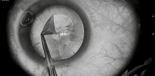
A small corneal incision, typically around two to three millimeters, provides access to the anterior chamber, and a continuous curvilinear capsulorhexis creates a smooth, round opening in the anterior lens capsule through which the cataract will be removed.
Hydrodissection separates the lens from its capsule with a fluid wave, and the phacoemulsification probe then uses ultrasonic energy to fragment the lens nucleus into small pieces that are simultaneously aspirated from the eye.
Once the lens material is fully removed, an intraocular lens, folded to fit through the small incision, is inserted into the empty capsular bag and unfolds into position, restoring the eye’s focusing power in place of the natural lens.
Why the Small Incision Matters
A properly constructed phacoemulsification incision is self-sealing, relying on the natural architecture of a carefully angled corneal wound rather than sutures to remain watertight, which is a major reason for the procedure’s fast recovery and minimal induced astigmatism.
This is in direct contrast to the larger incision required for extracapsular extraction, which needed multiple sutures to close, induced more astigmatism, and required a considerably longer recovery before vision stabilized.
The combination of a small, self-sealing incision and a foldable intraocular lens is what allows the entire procedure to typically be completed in well under half an hour, usually under topical or minimal local anesthesia rather than a full general anesthetic.
Patients are often surprised by how brief the procedure and the initial recovery period feel, and setting that expectation in advance helps replace any lingering fear based on older accounts of cataract surgery with a more accurate picture of what to expect today.
All-fit Slit-Lamp Adapter
Record and share exactly what you see at the slit lamp. One adapter fits any slit lamp or surgical microscope — and any smartphone.
From Choroida — the team behind this siteThe Capsulorhexis
Creating a smooth, continuous, appropriately sized circular opening in the anterior capsule is one of the more technically important steps in the entire procedure, since an irregular or radially extending tear can complicate the rest of the surgery significantly.
A well-centered, correctly sized capsulorhexis also helps ensure the intraocular lens is well-centered and stable within the capsular bag once inserted, which has direct implications for the patient’s final visual outcome.
Femtosecond laser-assisted cataract surgery, discussed in its own dedicated article on this site, automates this and several other steps of the procedure using laser rather than purely manual technique.
Energy and Fluidics
Modern phacoemulsification machines allow surgeons to fine-tune the balance between ultrasonic energy delivery, fluid inflow, and aspiration, a balance often referred to as fluidics, to safely and efficiently remove lens material while maintaining a stable anterior chamber throughout the case.
Excessive ultrasonic energy or poorly managed fluidics can stress the corneal endothelium, discussed in relation to specular microscopy in its own dedicated article on this site, which is part of why minimizing energy use, particularly in eyes with reduced endothelial reserve, is an active surgical consideration rather than an afterthought.
Denser, harder cataracts generally require more ultrasonic energy to fragment, which is one of several reasons a more advanced, longstanding cataract can carry somewhat higher surgical risk than one removed earlier in its natural progression.
Complications
Posterior capsule rupture is one of the more significant intraoperative complications, since it can complicate intraocular lens placement and increase the risk of further complications including vitreous loss and, later, retinal detachment.
Endophthalmitis, though rare given modern sterile technique and perioperative antibiotic prophylaxis, remains the most serious potential postoperative infectious complication and is treated as a genuine surgical emergency when it occurs.
Posterior capsule opacification, discussed in relation to YAG laser capsulotomy in its own dedicated article on this site, is the most common longer-term consequence of successful surgery, addressed easily with a brief outpatient laser procedure when it develops.
Cystoid macular edema and, rarely, retinal detachment can also follow otherwise uncomplicated surgery, which is why routine postoperative follow-up continues for several weeks even in an eye that appears to be recovering entirely normally.


Document what you see
Two smartphone imaging tools built for everyday clinic use — one for the slit lamp, one for the fundus.
From Choroida — the team behind this siteReferences
- American Academy of Ophthalmology. Basic and Clinical Science Course, Section 11: Lens and Cataract.
- Kelman CD. Phaco-emulsification and aspiration: a new technique of cataract removal. American Journal of Ophthalmology.
- Davis G. The evolution of cataract surgery. Missouri Medicine.
Test yourself
A few questions straight from this article.
-
In phacoemulsification, what is the ultrasonic energy used to do?
The phaco probe breaks the nucleus into small pieces that are aspirated at the same time, which is what allows the lens to leave through a small incision. -
What is the typical size of the corneal incision used for phacoemulsification?
An incision of roughly two to three millimetres gives access to the anterior chamber and is small enough to seal itself without sutures. -
What does hydrodissection achieve during phacoemulsification?
A fluid wave is injected to free the lens from the capsule before ultrasonic fragmentation of the nucleus begins. -
Why does a properly built phacoemulsification incision stay watertight without sutures?
The wound relies on the natural architecture of an angled corneal incision, which is a major reason recovery is quick and induced astigmatism minimal. -
Compared with phacoemulsification, extracapsular cataract extraction required what?
The older technique removed the nucleus intact through a much larger wound closed with multiple sutures, inducing more astigmatism and a longer recovery. -
Why does a well-centred, correctly sized capsulorhexis matter for the visual outcome?
A smooth, appropriately sized capsular opening helps the implanted lens sit centred and stable in the capsular bag, which bears directly on the final result. -
Excessive ultrasonic energy or poorly managed fluidics particularly stresses which structure?
The endothelium bears the brunt of excess energy and unstable fluidics, which is why energy use is minimised especially in eyes with reduced endothelial reserve. -
Why does a dense, longstanding cataract carry somewhat higher surgical risk?
Harder nuclei demand more ultrasonic energy to break up, which is one reason removing a cataract earlier in its course is generally less risky. -
What is the most common longer-term consequence of successful phacoemulsification?
Opacification of the posterior capsule is the commonest late consequence of otherwise successful surgery and is dealt with by a brief outpatient laser procedure. -
Which complication after cataract surgery is treated as a genuine surgical emergency?
Endophthalmitis is rare with modern sterile technique and antibiotic prophylaxis but remains the most serious postoperative infection and is handled as an emergency.

Writes and reviews the clinical articles on Educate. Every post is checked against peer-reviewed sources before it goes live.


