
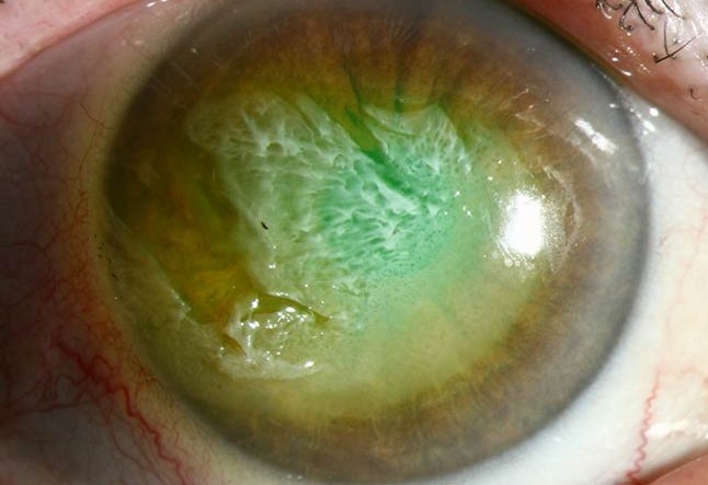
A thermal burn to the ocular surface occurs less often than a chemical burn but shares much of the same emergency assessment and limbal stem cell risk, while differing in one important respect: the reflexive, extremely fast blink response usually protects the cornea itself from prolonged heat exposure, so damage is often concentrated on the eyelids and periocular skin, with the globe affected more by brief flash exposure than by the sustained contact typical of a severe chemical burn.
Mechanisms of Injury
- Direct flame or fire exposure, from house fires, explosions, or industrial accidents, often affecting the eyelids and periocular skin more than the globe given the protective blink reflex
- Hot liquid or grease splash
- Contact with a heated object, such as a curling iron or hot metal, a relatively common household mechanism, particularly in children
- Steam exposure
- Welding and industrial flash burns, which combine thermal and, in some cases, ultraviolet injury
Why the Blink Reflex Matters
The blink reflex is extremely fast, typically completing within a fraction of a second of a threatening stimulus, and this speed usually limits direct corneal thermal exposure to a very brief period even during a significant burn event, in contrast to a chemical burn, where the reflex closes the lids but does not stop the chemical from continuing to react with whatever tissue it has already contacted.
This is why, in many thermal injuries, the eyelids bear the brunt of the burn while the globe itself, though still requiring careful assessment, is often less severely affected than the periocular skin would suggest.
All-fit Slit-Lamp Adapter
Record and share exactly what you see at the slit lamp. One adapter fits any slit lamp or surgical microscope — and any smartphone.
From Choroida — the team behind this siteAssessment
- Assessment of the eyelid skin burn depth and extent, using standard burn classification (superficial, partial-thickness, full-thickness), since this affects both immediate management and the risk of later lid malposition
- Careful examination of the globe itself, including the cornea, conjunctiva, and limbus, for any area of thermal injury, whitening, or epithelial loss
- Assessment of limbal involvement specifically, since limbal stem cell injury, even from a brief thermal exposure, carries the same long-term risk of limbal stem cell deficiency as seen with chemical burns (see limbal stem cell deficiency)
- Evaluation for associated injuries, including inhalation injury in a fire setting, which takes priority in the overall trauma assessment
Immediate Management
- Copious irrigation is less central to thermal burn management than to chemical burns, since there is no ongoing chemical reaction to dilute, though initial cooling and removal of any residual hot material (such as embedded debris) is appropriate
- Careful debridement of any loose, clearly nonviable tissue
- Topical antibiotic ointment and lubrication for corneal or conjunctival epithelial defects
- A bandage contact lens or, for more significant epithelial loss, amniotic membrane, similar in principle to management of other significant epithelial defects
Managing Limbal Involvement
When examination shows limbal ischemia or significant limbal epithelial loss, the same staging and concern that applies to chemical burns applies here: greater circumferential limbal involvement predicts a higher risk of later limbal stem cell deficiency, chronic ocular surface failure, and corneal vascularization, and these eyes need closer follow-up and earlier consideration of surface-supporting interventions such as amniotic membrane transplantation.
Managing Eyelid Injury
- Superficial eyelid burns are managed with topical antibiotic ointment and wound care
- Deeper, full-thickness eyelid burns carry a significant risk of later cicatricial eyelid malposition, including ectropion and lagophthalmos, which can secondarily expose and damage the cornea even after the primary burn has healed
- Close follow-up through the healing and scarring process is needed to identify and address early lid malposition before it causes exposure keratopathy, sometimes requiring later reconstructive surgery
Long-Term Follow-Up
Patients with significant thermal burns to the periocular region need follow-up extending well beyond the initial acute injury, both for the ocular surface (limbal stem cell function, dry eye, corneal clarity) and for the eyelids (position, closure, and any need for scar release or reconstruction).
Prognosis
Isolated thermal injury without significant limbal involvement generally has a good visual prognosis, given the protective effect of the blink reflex on the globe itself.
The main long-term threats to vision are significant limbal stem cell loss, when it does occur, and secondary exposure keratopathy from cicatricial eyelid malposition, both of which require ongoing surveillance well after the acute burn has healed.


Document what you see
Two smartphone imaging tools built for everyday clinic use — one for the slit lamp, one for the fundus.
From Choroida — the team behind this siteReferences
- Kuckelkorn R, Kottek A, Reim M. Intraocular complications after severe chemical burns–incidence and surgical treatment. Ophthalmologica. 1994;208:294-301.
- Fish R, Davidson RS. Management of ocular thermal and chemical injuries, including amniotic membrane therapy. Curr Opin Ophthalmol. 2010;21:317-321.
- Dua HS, King AJ, Joseph A. A new classification of ocular surface burns. Br J Ophthalmol. 2001;85:1379-1383.
- Wagoner MD. Chemical injuries of the eye: current concepts in pathophysiology and therapy. Surv Ophthalmol. 1997;41:275-313.
