The retinal artery occlusion workup is, in practice, a stroke workup performed in a patient who happens to present to an eye clinic.
Also, central and branch retinal artery occlusions are ischemic events in the territory of the internal carotid artery.
The patient is also at risk of a cerebral stroke in the days and weeks that follow.
Also, the ophthalmologist who recognizes this link should start the evaluation on the same day.
Retinal Artery Occlusion Workup: Why Treat It as a Stroke
The central retinal artery is a branch of the ophthalmic artery, which arises from the internal carotid artery.
Embolism from the carotid, the aortic arch, or the heart is also the commonest mechanism, and other causes include arteritis and hypercoagulable states.
Studies of patients with central retinal artery occlusion show that a substantial proportion have silent cerebral infarcts on diffusion-weighted imaging at presentation.
In addition, the American Heart Association has issued a statement. It advises treating central retinal artery occlusion as an acute ischemic stroke (see central retinal artery occlusion).
First Steps in the Retinal Artery Occlusion Workup
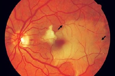
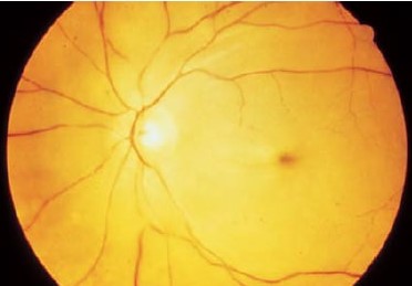
- Confirm the diagnosis with the history and the examination: sudden painless loss of vision, a relative afferent pupillary defect, a pale retina with a cherry-red spot, and attenuated arterioles
- Look for emboli in the retinal arteries (see Hollenhorst plaque)
- Check for a cilioretinal artery that may spare the macula (see cilioretinal artery occlusion)
- Ask about headache, jaw claudication, scalp tenderness, weight loss, and polymyalgia, which suggest giant cell arteritis
- Measure the blood pressure, pulse rate, and rhythm
- Send the patient on the same day to an emergency department or a stroke unit with experience in this evaluation
Fundus Explorer Pro
Photograph the retinal findings described here with the phone already in your pocket — 22 D optics and built-in illumination in one handheld unit.
From Choroida — the team behind this siteGiant Cell Arteritis
In patients older than 50, the erythrocyte sedimentation rate, the C-reactive protein.
Check the platelet count at once.
Arteritic occlusion is uncommon, but it is a medical emergency, since the other eye is at high risk without steroid treatment.
If the suspicion is high, begin high-dose corticosteroids without waiting for temporal artery biopsy, and arrange the biopsy promptly (see arteritic anterior ischemic optic neuropathy).
Retinal Artery Occlusion Workup: Investigations for the Source
- Brain imaging: MRI with diffusion-weighted sequences, or CT if MRI is unavailable
- Vascular imaging: carotid duplex, CT angiography, or MR angiography of the neck and the head
- Cardiac evaluation: an electrocardiogram, prolonged rhythm monitoring for atrial fibrillation, and an echocardiogram
- Blood tests: glucose, lipids, blood count, and inflammatory markers
In young patients, or in those without a clear source, extended tests for hypercoagulability, vasculitis, endocarditis, drug use, migraine, and inherited conditions are considered.
Acute Treatment
There is no proven treatment that restores vision in most patients.
Ocular massage, anterior chamber paracentesis, and lowering of the intraocular pressure have been used, but the evidence for benefit is weak.
Researchers have studied intravenous thrombolysis within a few hours of onset.
Results are mixed, so its use depends on local protocols and the time window.
Some centers consider hyperbaric oxygen for selected patients with early presentation.
Retinal Artery Occlusion Workup: Secondary Prevention
The main benefit comes from preventing the next event.
- Antiplatelet therapy, usually starting once doctors exclude hemorrhage
- Statin therapy and control of blood pressure and diabetes
- Intervention for symptomatic carotid stenosis, as for other carotid territory events
- Anticoagulation for atrial fibrillation
- Smoking cessation and lifestyle advice
Patients should also have follow-up by a stroke or vascular medicine service.
Inform the eye clinic of the results.
Explain that the loss of vision is usually permanent, and that the aim of the evaluation is to protect the patient from a more serious event.
Timing and Logistics
The first hours matter, because the stroke risk is highest in the first days, and delays in referral reduce the options for treatment and secondary prevention.
A written referral that states the diagnosis as retinal artery occlusion, and asks for a stroke-pathway evaluation, speeds the process.
If emergency services are not close, the clinic should call ahead and send a copy of the findings with the patient.
Branch and Cilioretinal Occlusions
A branch retinal artery occlusion usually causes a sectoral visual field defect.
The evaluation is the same as for central occlusion.
A cilioretinal artery occlusion in an eye with nonarteritic ischemic optic neuropathy or vein occlusion may have a different cause. The stroke evaluation is still indicated.
Transient monocular vision loss, or amaurosis fugax, needs a similar evaluation, even though the retina looks normal (see amaurosis fugax).
Communicating With the Patient
Explain that the eye has had a stroke. Add that the visual loss is unlikely to improve, and that the main risk now is a stroke in the brain.
Make sure that the patient understands the warning signs of a brain stroke, such as weakness, speech difficulty, or facial droop, and knows to call emergency services.
Follow the patient for neovascular complications, including rubeosis, which can appear in the weeks after the event.


Document what you see
Two smartphone imaging tools built for everyday clinic use — one for the slit lamp, one for the fundus.
From Choroida — the team behind this siteReferences
- Hayreh SS, Zimmerman MB. Central retinal artery occlusion: visual outcome. Am J Ophthalmol. 2005;140:376-391.
- Mac Grory B, Schrag M, Biousse V, et al. Management of central retinal artery occlusion: a scientific statement from the American Heart Association. Stroke. 2021;52:e282-e294.
- Hayreh SS. Central retinal artery occlusion. Indian J Ophthalmol. 2018;66:1684-1694.
- American Academy of Ophthalmology. Basic and Clinical Science Course, Section 12: Retina and Vitreous. AAO; current edition.

Writes and reviews the clinical articles on Educate. Every post is checked against peer-reviewed sources before it goes live.