
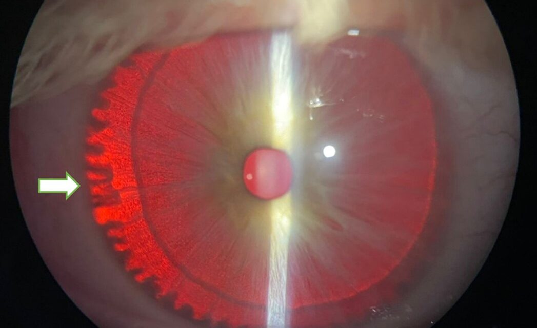
Iris transillumination defects are areas where the normally light-blocking posterior iris pigment epithelium has thinned or been lost, and because different underlying conditions produce transillumination defects in characteristically different locations and patterns, this simple bedside technique, shining a focused light through the pupil or sclera and watching where light escapes through the iris itself, can meaningfully narrow the differential diagnosis in seconds.
How Transillumination Works
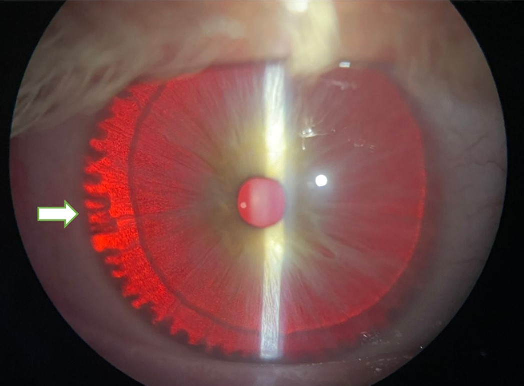
The technique involves directing a focused light source, either through the pupil (retroillumination) or through the sclera, and observing the iris from the front: where the posterior pigment epithelium is intact and healthy, it blocks the transmitted light effectively, while areas of pigment epithelial thinning or loss allow light to pass through, appearing as a bright red or orange glow against the surrounding normally dark iris.
Pattern-Based Differential Diagnosis
Pigment Dispersion Syndrome
Produces characteristic mid-peripheral, spoke-like or radial transillumination defects, resulting from mechanical rubbing of the posterior iris pigment epithelium against the anterior zonular fibers as the iris flexes with normal pupillary movement, a mechanism specific enough that this pattern is considered classic for the condition and directly relevant to its associated pigmentary glaucoma risk.
Albinism
Produces diffuse transillumination throughout the entire iris, reflecting the generalized reduction in ocular pigmentation characteristic of albinism, in clear contrast to the more localized, patterned defects seen in pigment dispersion syndrome or trauma, and is typically accompanied by other characteristic ocular findings, including foveal hypoplasia and nystagmus (see albinism and foveal hypoplasia).
Trauma
Produces focal, often irregular transillumination defects at the specific site of prior injury, sometimes corresponding to a visible iris sphincter tear or other structural disruption, and typically unilateral and asymmetric with the fellow eye, distinguishing it from the more symmetric, bilateral patterns of pigment dispersion syndrome or albinism.
Pseudoexfoliation Syndrome
Produces transillumination defects typically concentrated at the pupillary margin, related to chronic mechanical rubbing between the iris and the pseudoexfoliative material deposited on the anterior lens capsule, a distribution pattern distinct from the mid-peripheral pattern of pigment dispersion syndrome.
Herpetic Iridocyclitis
Can produce patchy, sector-shaped transillumination defects following an episode of herpes simplex or herpes zoster-related anterior segment inflammation, reflecting focal iris ischemia and pigment epithelial damage from the inflammatory process itself.
All-fit Slit-Lamp Adapter
Record and share exactly what you see at the slit lamp. One adapter fits any slit lamp or surgical microscope — and any smartphone.
From Choroida — the team behind this siteClinical Significance
- Confirming pigment dispersion syndrome supports appropriate monitoring for pigmentary glaucoma, given the meaningful proportion of affected patients who go on to develop elevated intraocular pressure
- Recognizing a traumatic pattern supports a more complete assessment for other, sometimes more visually threatening, consequences of the same injury, including angle recession and lens subluxation (see angle recession glaucoma)
- Diffuse transillumination in an infant or young child prompts full evaluation for albinism, including assessment of visual acuity, nystagmus, and foveal development
- A sector-shaped pattern following a red eye episode supports a herpetic cause even when the acute presentation has already resolved and the patient no longer recalls the details clearly
Examination Technique
Both transillumination methods, retroillumination through the pupil and scleral transillumination, are performed at the slit lamp with the room lights dimmed for optimal visualization, and both eyes should be examined and compared, since bilaterality, symmetry, and the specific pattern observed all contribute meaningfully to the differential diagnosis.
Clinical Value
Iris transillumination defects are a straightforward, always-available bedside finding that, evaluated with attention to pattern and distribution rather than simply noted as present or absent, meaningfully narrows the differential diagnosis for a range of anterior segment conditions, from a common, glaucoma-relevant pigment dispersion process to a distinctly rarer, systemic pigmentary disorder.


Document what you see
Two smartphone imaging tools built for everyday clinic use — one for the slit lamp, one for the fundus.
From Choroida — the team behind this siteReferences
- Campbell DG. Pigmentary dispersion and glaucoma. A new theory. Arch Ophthalmol. 1979;97:1667-1672.
- Summers CG. Albinism: classification, clinical characteristics, and recent findings. Optom Vis Sci. 2009;86:659-662.
- Ritch R. A unification hypothesis of pigment dispersion syndrome. Trans Am Ophthalmol Soc. 1996;94:381-405.
- Liesegang TJ. Herpes zoster ophthalmicus natural history, risk factors, clinical presentation, and morbidity. Ophthalmology. 2008;115:S3-12.
