Fellow eye retinal detachment risk is a question every retinal surgeon faces when a patient presents with a detachment in one eye.
The other eye has a higher chance of detaching than the average eye, and the decision about whether to treat it before it happens depends on what is seen.
The evidence is limited, so judgment and patient preference matter.
How High Is the Fellow Eye Retinal Detachment Risk?
Published series suggest that about 5 to 10 percent of patients with a rhegmatogenous retinal detachment in one eye will develop a detachment in the other eye.
The risk is higher in some groups and lower in others, and it depends on the time period and the type of detachment.
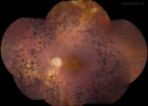
Patients with lattice degeneration in the first eye, with a history of cataract surgery, or with high myopia are at the top of the range.
Folk and colleagues studied the fellow eyes of patients with detachment attributed to lattice degeneration, and found a significant rate of bilateral disease (see the references).
Fellow Eye Retinal Detachment Risk: Factors That Raise the Risk
- Lattice degeneration in the fellow eye, especially with atrophic holes or tears (see lattice degeneration)
- High myopia, with long axial length (see retinal detachment in high myopia)
- Pseudophakia or aphakia, after cataract surgery, particularly with complications (see pseudophakic retinal detachment)
- Family history of detachment
- Connective tissue disorders, such as Stickler syndrome, in which the risk is very high (see Stickler syndrome)
- Trauma that affected both eyes
- Symptomatic posterior vitreous detachment with new tears
- Giant retinal tear in the first eye, a strong risk factor for the fellow eye (see giant retinal tear)
Fundus Explorer Pro
Photograph the retinal findings described here with the phone already in your pocket — 22 D optics and built-in illumination in one handheld unit.
From Choroida — the team behind this siteWhat to Examine
A dilated examination with scleral depression of the fellow eye is part of the evaluation of every patient with a detachment (see scleral depression).
The examiner should look for lattice, tears, holes, vitreous traction, and areas of vitreoretinal adhesion.
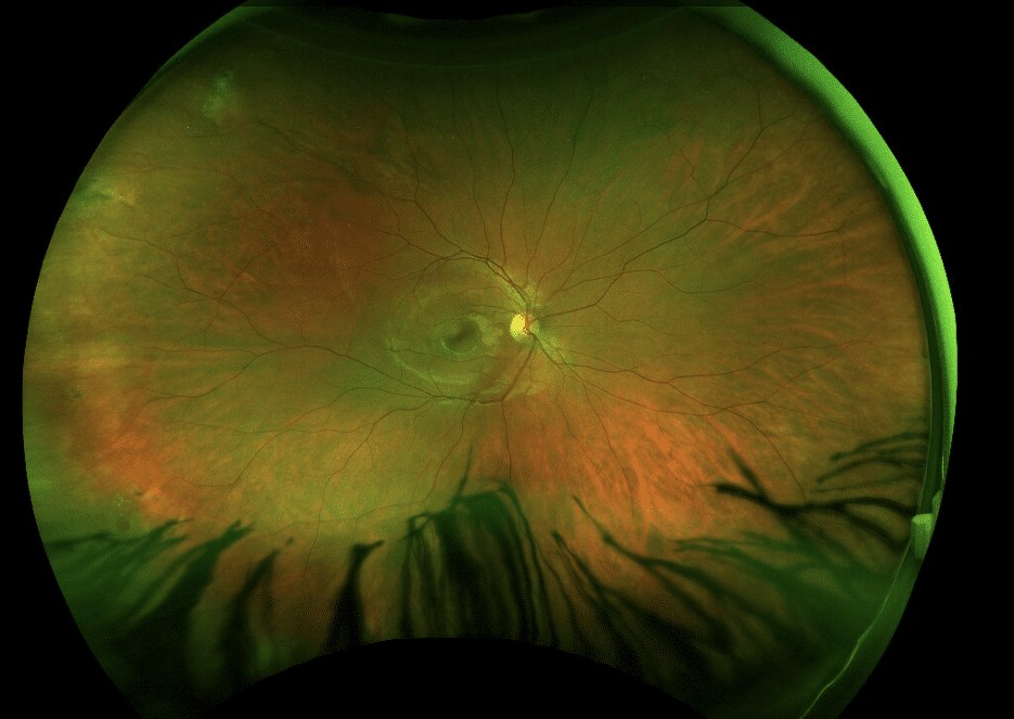
Widefield imaging can document findings and help in follow-up (see ultra-widefield retinal imaging).
Look at the condition of the vitreous, since a posterior vitreous detachment lowers the risk from lattice but may cause new tears.
When to Treat Prophylactically
- Symptomatic tears or horseshoe tears: treat promptly, with laser retinopexy or cryotherapy (see laser retinopexy)
- Lattice with atrophic holes or tears, in a patient with a first-eye detachment: many surgeons offer prophylaxis
- Stickler syndrome: prophylaxis with laser or cryotherapy, often 360 degrees, is commonly advised
- High myopia with extensive lattice and a family history: consider treatment
- Asymptomatic lattice without breaks: often observed, as evidence for benefit is weak
The Cochrane review concluded that there is not enough evidence to prove that prophylactic treatment prevents detachment, but that it is considered reasonable in high-risk situations (see the references).
Fellow Eye Retinal Detachment Risk: Risks of Prophylaxis
- Failure to prevent detachment, and new breaks at the edge of treatment
- Complications of laser or cryotherapy, such as pain, hemorrhage, and rarely macular pucker
- Overtreatment of lesions that would never have caused problems
Tell patients that prophylaxis lowers the risk, but does not eliminate it, and that they must still watch for symptoms.
Fellow Eye Retinal Detachment Risk: Follow-Up and Education
- Review the fellow eye at intervals after the first detachment, for example at 3 to 6 months and then yearly
- Provide a written list of warning symptoms, with a number to call
- Encourage the patient to report new floaters or flashes at once
- Take extra care before cataract surgery in either eye, and discuss the risk
Patients who have had one detachment are often highly motivated to avoid another, and clear information helps.
Counselling
Explain that the fellow eye has a higher risk, but that most do not detach, and that examination and treatment can help.
Discuss the options in terms of benefits and risks, and allow the patient to take part in the decision.
Talking About Risk With the Patient
- Use plain numbers, such as “about one in ten”, and avoid ambiguous words
- Explain that the risk is higher for certain features, and that we know what to look for
- Describe what prophylaxis involves, how long it takes, and the common side effects
- Check that the patient understands that the treatment does not give complete protection
Shared decisions improve satisfaction, and patients often choose treatment when they have seen a detachment in the first eye.
After a Detachment in the First Eye
The first eye will need follow-up too, and recurrence is possible, especially if PVR develops.
The patient may be anxious about both eyes, and may need extra support and practical advice about warning signs.
Review the retina periphery at each visit, and make sure that both eyes are covered by the plan.
For the latest studies, see the PubMed results for fellow eye retinal detachment risk.


Document what you see
Two smartphone imaging tools built for everyday clinic use — one for the slit lamp, one for the fundus.
From Choroida — the team behind this siteReferences
- Folk JC, Arrindell EL, Klugman MR. The fellow eye of patients with phakic lattice retinal detachment. Ophthalmology. 1989;96:72-79.
- Wilkinson CP. Interventions for asymptomatic retinal breaks and lattice degeneration for preventing retinal detachment. Cochrane Database Syst Rev. 2014;9:CD003170.
- Ryan SJ, Schachat AP, Wilkinson CP, et al, eds. Retina. 6th ed. Elsevier; 2018.
- American Academy of Ophthalmology. Basic and Clinical Science Course, Section 12: Retina and Vitreous. AAO; current edition.

Writes and reviews the clinical articles on Educate. Every post is checked against peer-reviewed sources before it goes live.