
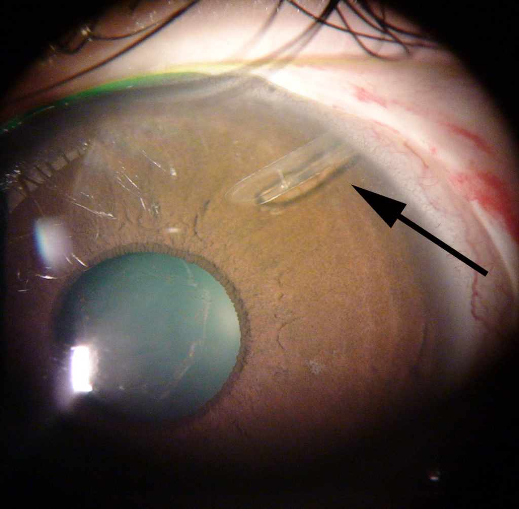
Glaucoma drainage device surgery, commonly called a tube shunt, uses an implanted silicone tube connected to an external plate to drain aqueous from the anterior chamber into a reservoir formed by fibrous tissue around the plate.
It represents a fundamentally different engineering solution from trabeculectomy: instead of relying on a scleral flap and a conjunctival bleb, drainage is routed through a fixed tube to a controlled, distant collection point.
That structural difference is what makes tube shunts the preferred option in exactly the situations where trabeculectomy tends to fail.
Several implant designs exist, differing mainly in plate size, material, and whether they include a built-in valve, and the choice among them is guided by surgeon experience and the specific clinical scenario rather than one design being universally superior.
How It Works
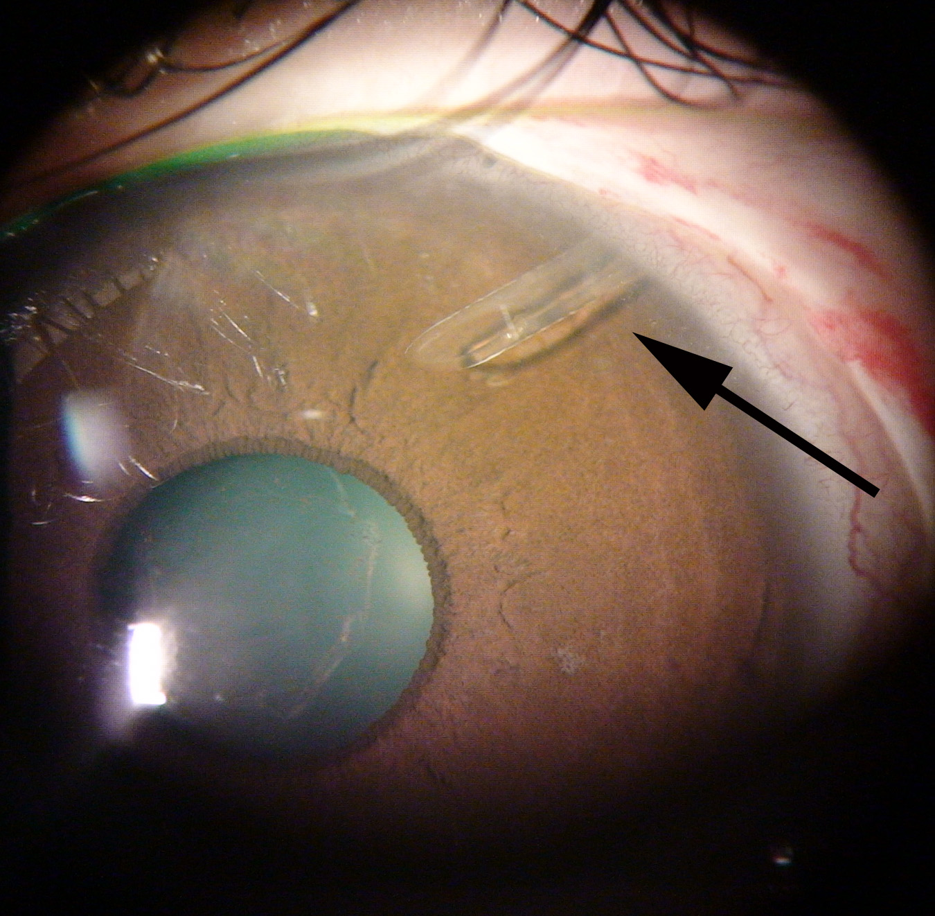
A tube is inserted into the anterior chamber, or occasionally the pars plana in eyes without adequate anterior chamber access, and connected to a plate sutured to the sclera, usually in a posterior quadrant well away from the visual axis.
Aqueous flows through the tube to the plate, where the body forms a fibrous capsule, or bleb, around the plate itself, and it is the resistance of this capsule that ultimately governs the pressure achieved rather than any valve mechanism within the tube itself in most designs.
Non-valved devices like the Baerveldt implant require a temporary tube occlusion or a restrictive suture in the early postoperative period to prevent immediate overdrainage, since the capsule has not yet formed enough resistance to regulate flow on its own.
Valved devices like the Ahmed implant include a mechanical valve intended to reduce the risk of early hypotony, though the valve’s flow resistance is only part of the pressure picture once the fibrous capsule matures.
A direct comparison between the two major device families, the Ahmed Versus Baerveldt Study, found broadly similar long-term pressure control but a different early complication profile, with the valved device showing less early hypotony and the non-valved device sometimes achieving lower pressures once the capsule matured.
Indications
- Glaucoma that has failed one or more prior trabeculectomies, since scarring from previous filtering surgery makes another trabeculectomy less likely to succeed
- Neovascular glaucoma, where trabeculectomy carries a particularly high failure and complication rate
- Uveitic glaucoma, where ongoing inflammation similarly predisposes trabeculectomy blebs to scar
- Eyes with extensive conjunctival scarring from prior surgery that would compromise a trabeculectomy bleb
- As an increasingly common primary option in some practice patterns, based on comparative trial data discussed below
All-fit Slit-Lamp Adapter
Record and share exactly what you see at the slit lamp. One adapter fits any slit lamp or surgical microscope — and any smartphone.
From Choroida — the team behind this siteTube Versus Trabeculectomy
The Tube Versus Trabeculectomy Study, a major randomized comparison in eyes with prior ocular surgery, found tube shunts had a lower failure rate than trabeculectomy with mitomycin-C over long-term follow-up in that specific population.
This shifted practice meaningfully toward earlier use of tube shunts in eyes with a surgical history, though trabeculectomy retains an edge in achieving very low target pressures in appropriately selected, surgically naive eyes.
Neither procedure has rendered the other obsolete, and the choice remains individualized based on prior surgical history, conjunctival status, and the specific target pressure needed.
Complications
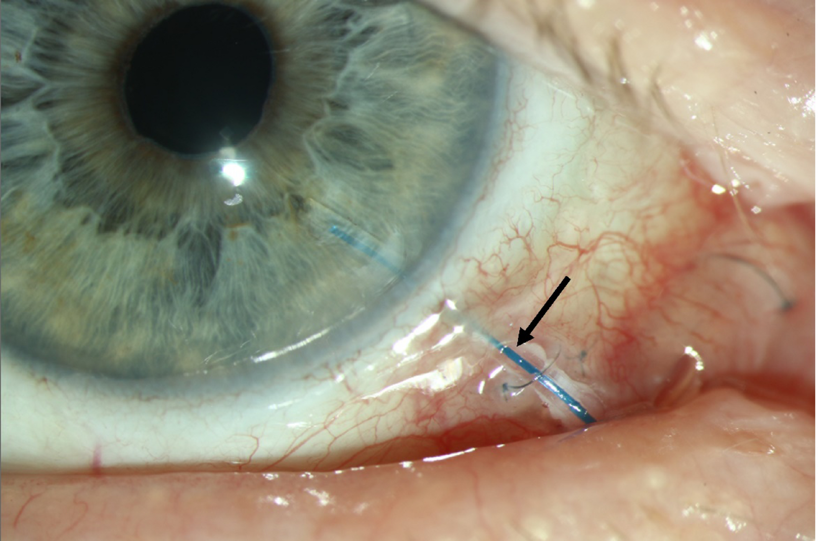
- Tube exposure through the conjunctiva, which creates an infection risk and typically requires surgical revision with a patch graft
- Corneal endothelial decompensation from tube-cornea touch or chronic proximity, particularly with anteriorly positioned tubes
- Diplopia from the plate’s bulk affecting extraocular muscle function, especially with larger plate designs
- Hypotony, most classically an early risk with non-valved devices before the fibrous capsule has matured enough to regulate flow
- Tube malposition or obstruction, sometimes from iris, vitreous, or fibrin plugging the tube’s internal opening
Postoperative Course
Early follow-up focuses on confirming appropriate pressure control and watching for the transient pressure spike that can occur as the fibrous capsule around the plate matures over the first several weeks, sometimes called the hypertensive phase.
This hypertensive phase is a recognized, usually self-limited part of the normal healing course with non-valved devices, and it typically responds to temporary medical therapy rather than requiring further surgery.
Long-term monitoring continues to track both intraocular pressure and the tube’s physical position relative to the cornea and conjunctiva, since late tube exposure or corneal complications can develop years after otherwise successful surgery.
Endothelial cell density is worth checking periodically in eyes with an anteriorly positioned tube, since gradual endothelial cell loss from chronic tube-cornea proximity can progress silently over years before becoming clinically apparent as corneal edema.


Document what you see
Two smartphone imaging tools built for everyday clinic use — one for the slit lamp, one for the fundus.
From Choroida — the team behind this siteReferences
- Gedde SJ, Schiffman JC, Feuer WJ, et al. Treatment outcomes in the Tube Versus Trabeculectomy Study after five years of follow-up. American Journal of Ophthalmology.
- American Academy of Ophthalmology. Basic and Clinical Science Course, Section 10: Glaucoma.
- Christakis PG, Kalenak JW, Zurakowski D, et al. The Ahmed Versus Baerveldt Study: results after one year of follow-up. Ophthalmology.
Test yourself
A few questions straight from this article.
-
In glaucoma drainage device surgery, where does the implanted tube carry aqueous from the anterior chamber?
The tube routes aqueous to an external plate sutured to the sclera, where the body forms a fibrous capsule that serves as the reservoir, rather than to a scleral flap and conjunctival bleb. -
In most glaucoma drainage device designs, what mainly determines the intraocular pressure finally achieved?
Pressure is governed by the resistance of the fibrous capsule that forms around the plate, not by any valve mechanism inside the tube in most designs. -
Why does a non-valved implant such as the Baerveldt need temporary tube occlusion in the early postoperative period?
Before the capsule has formed enough resistance to regulate flow, a temporary tube occlusion or restrictive suture is needed to prevent immediate overdrainage. -
The Ahmed Versus Baerveldt Study compared valved and non-valved implants. What did it find about early complications?
Long-term pressure control was broadly similar, but the early complication profile differed: less early hypotony with the valved device, and sometimes lower pressures with the non-valved device once the capsule matured. -
The Tube Versus Trabeculectomy Study enrolled eyes with prior ocular surgery. What was its long-term finding?
In that specific population of eyes with previous ocular surgery, tube shunts had a lower failure rate over long-term follow-up, which shifted practice toward earlier tube use. -
In which eyes does trabeculectomy still hold an advantage over a glaucoma drainage device?
Trabeculectomy retains an edge in reaching very low target pressures in appropriately selected, surgically naive eyes; the other scenarios listed favour a tube. -
Which glaucoma type is cited as carrying a particularly high trabeculectomy failure rate, favouring a tube shunt instead?
Neovascular glaucoma is listed as a tube shunt indication because trabeculectomy carries a particularly high failure and complication rate in these eyes. -
A glaucoma drainage device tube becomes exposed through the overlying conjunctiva. What does this typically require?
Tube exposure creates an infection risk and typically requires surgical revision with a patch graft rather than observation. -
Weeks after tube shunt surgery the pressure rises transiently as the capsule matures. How is this hypertensive phase usually managed?
The hypertensive phase is a recognised and usually self-limited part of normal healing as the capsule around the plate matures, and it typically settles with temporary medical therapy. -
Why is endothelial cell density checked periodically in eyes with an anteriorly positioned drainage tube?
Chronic tube-cornea proximity causes gradual endothelial cell loss that progresses silently for years before presenting clinically as corneal edema.

Writes and reviews the clinical articles on Educate. Every post is checked against peer-reviewed sources before it goes live.


