
Vitreomacular adhesion describes the earliest, mildest point on a spectrum of vitreomacular interface disease, and the entire clinical value of separating this specific stage out from the more advanced, symptomatic stages that can follow it lies in the different management path it implies: watchful monitoring rather than the surgical consideration warranted once adhesion progresses to genuine traction.
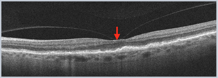
The Vitreomacular Interface Spectrum
As part of the normal aging process, the vitreous gel gradually separates from the retina in a process called posterior vitreous detachment, and this separation does not always occur uniformly or completely, sometimes leaving persistent focal attachment specifically at the fovea even as the vitreous separates more completely elsewhere across the posterior pole.
This persistent focal attachment is what defines vitreomacular adhesion, and it represents a distinct, earlier stage from vitreomacular traction, in which the same persistent attachment has progressed to the point of actually distorting the underlying foveal anatomy.
Vitreomacular Adhesion Versus Vitreomacular Traction
The International Vitreomacular Traction Study Group established standardized criteria, based on optical coherence tomography, for distinguishing these related but clinically distinct stages.
- Vitreomacular adhesion describes persistent vitreofoveal attachment without any associated distortion of the underlying retinal architecture, essentially a structural finding without functional consequence
- Vitreomacular traction describes the same persistent attachment, but now with associated distortion of the foveal contour or underlying retinal architecture, reflecting genuine mechanical pulling on the retina significant enough to alter its normal structure
This distinction matters directly for management, since vitreomacular adhesion alone, without associated traction or distortion, is generally asymptomatic and does not require intervention, while vitreomacular traction causing significant symptoms may eventually warrant treatment.
Fundus Explorer Pro
Photograph the retinal findings described here with the phone already in your pocket — 22 D optics and built-in illumination in one handheld unit.
From Choroida — the team behind this siteClinical Significance
- Vitreomacular adhesion is a common finding on optical coherence tomography, particularly in older patients undergoing macular imaging for another reason, and its presence alone, without accompanying traction or macular distortion, is not itself considered pathologic
- Vitreomacular adhesion can resolve spontaneously as the vitreous eventually completes its separation from the macula, or it can persist and, in a subset of patients, progress to symptomatic vitreomacular traction over time
- Vitreomacular adhesion is also relevant as a precursor consideration in full-thickness macular hole formation, since persistent focal vitreofoveal attachment is part of the mechanical process believed to contribute to some macular hole development (see lamellar versus full-thickness macular hole for the related discussion of hole formation and staging)
Diagnostic Evaluation
- Optical coherence tomography is the essential tool for identifying and precisely characterizing vitreomacular adhesion, directly visualizing the persistent vitreofoveal attachment and assessing whether any associated distortion of the foveal contour is present
- Careful clinical correlation with the patient’s visual symptoms, since adhesion without traction is typically asymptomatic, while the presence of new visual distortion or metamorphopsia should prompt closer assessment for progression to traction
Management
Observation
Isolated vitreomacular adhesion, without associated traction, distortion, or visual symptoms, is generally managed with observation alone, given its frequently benign, sometimes self-resolving natural history.
Monitoring for Progression
Periodic follow-up, including repeat optical coherence tomography imaging, is reasonable for patients with vitreomacular adhesion, particularly when there is any early or subtle evidence suggesting a trend toward developing traction, allowing earlier identification of progression should it occur.
Management if Progression to Symptomatic Traction Occurs
If vitreomacular adhesion progresses to symptomatic vitreomacular traction with significant visual impact, treatment options, including pharmacologic vitreolysis in appropriately selected cases or surgical vitrectomy for more extensive or non-resolving traction, are considered, following the broader management principles applied to symptomatic vitreomacular traction syndrome.
What the OCT shows
In vitreomacular adhesion, the posterior hyaloid remains attached to the macula over a defined width while the rest of the vitreous has separated, but the foveal contour is normal. In vitreomacular traction, the attachment pulls and distorts the contour, and may produce cysts or a hole. Measuring the width of the adhesion and checking for an epiretinal membrane helps to predict whether it will release by itself.
When to monitor
An asymptomatic adhesion with a normal foveal contour needs only observation, since many release spontaneously. Patients should check each eye with an Amsler grid, and report new distortion. If traction develops, see vitreoretinal traction syndrome for the treatment options.
Prognosis
Vitreomacular adhesion alone carries an excellent prognosis, and many cases resolve spontaneously over time as the natural process of posterior vitreous separation eventually completes without ever progressing to symptomatic traction.
The main clinical value of recognizing and correctly staging vitreomacular adhesion lies in appropriately reassuring patients with this isolated, asymptomatic finding while maintaining reasonable surveillance for the smaller subset who do progress to a stage warranting active treatment.


Document what you see
Two smartphone imaging tools built for everyday clinic use — one for the slit lamp, one for the fundus.
From Choroida — the team behind this siteReferences
- Duker JS, Kaiser PK, Binder S, et al. The International Vitreomacular Traction Study Group classification of vitreomacular adhesion, traction, and macular hole. Ophthalmology. 2013;120:2611-2619.
- Johnson MW. Posterior vitreous detachment: evolution and complications of its early stages. Am J Ophthalmol. 2010;149:371-382.e1.
- Steel DH, Lotery AJ. Idiopathic vitreomacular traction and macular hole: a comprehensive review of pathophysiology, diagnosis, and treatment. Eye (Lond). 2013;27 Suppl 1:S1-S21.
- Stalmans P, Benz MS, Gandorfer A, et al. Enzymatic vitreolysis with ocriplasmin for vitreomacular traction and macular holes. N Engl J Med. 2012;367:606-615.
