
Trabeculectomy is the classic filtering surgery for glaucoma, creating a new drainage pathway that lets aqueous humor bypass the eye’s own diseased outflow system and drain into a space under the conjunctiva.
It has remained the reference-standard glaucoma surgery for decades, and even as newer procedures have multiplied, it is still the benchmark against which those alternatives are measured for how much pressure lowering they can actually achieve.
That pressure-lowering power comes with a real complication profile, which is exactly why patient selection and postoperative bleb management matter as much as the operation itself.
The procedure dates back to the late 1960s, and while surgical instrumentation and antifibrotic adjuncts have evolved considerably since then, the core mechanism has remained essentially unchanged.
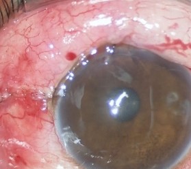
Surgical Principle
The surgeon creates a partial-thickness scleral flap, then fashions an opening into the anterior chamber beneath it, allowing aqueous to flow out from inside the eye into a space under the flap and, ultimately, under the conjunctiva.
The conjunctiva overlying this new drainage route forms a filtering bleb, a raised, fluid-filled area that becomes the visible hallmark of a successful trabeculectomy and the structure that subsequent care is built around.
The scleral flap is sutured back down at a tension calibrated to control the rate of aqueous outflow, since a flap that is too loose causes early postoperative hypotony and one that is too tight fails to lower pressure adequately.
Role of Antifibrotic Agents
Mitomycin-C or, less commonly, 5-fluorouracil is applied intraoperatively to reduce scarring at the surgical site, since the eye’s natural wound-healing response is the single biggest threat to a trabeculectomy’s long-term success.
Antifibrotic use lowered failure rates substantially compared to trabeculectomy without adjunctive treatment, but it also increases the risk of an overly thin, avascular bleb, which in turn raises the risk of late bleb leak and infection.
Balancing enough antifibrotic effect to prevent scarring against too much, which predisposes to a fragile bleb, is one of the central judgment calls in modern trabeculectomy technique.
The concentration, exposure time, and placement of the antifibrotic sponge are all adjusted based on the individual eye’s scarring risk, including prior ocular surgery, ethnicity, and age, since younger and more heavily pigmented eyes tend to scar more aggressively.
All-fit Slit-Lamp Adapter
Record and share exactly what you see at the slit lamp. One adapter fits any slit lamp or surgical microscope — and any smartphone.
From Choroida — the team behind this siteIndications
- Glaucoma with progressive optic nerve damage or visual field loss despite maximal tolerated medical therapy
- Glaucoma uncontrolled after laser trabeculoplasty or other less invasive interventions
- A target pressure low enough that medical therapy or minimally invasive glaucoma surgery is unlikely to achieve it reliably
- Patient factors such as medication intolerance or poor adherence that make long-term medical control impractical
Postoperative Bleb Management
Close early follow-up is essential, since the pressure trajectory in the first weeks after surgery determines whether laser suture lysis or releasable sutures need to be adjusted to fine-tune outflow.
A functioning bleb needs to be distinguished from a failing one at each visit: a diffuse, low, cystic bleb with a well-controlled pressure looks very different from an elevated, vascularized, scarring bleb that signals impending failure.
Bleb massage, needling with an adjunctive antifibrotic injection, or a return to the operating room may all be used to rescue a bleb that is beginning to fail from early scarring, depending on how far the process has progressed.
Complications
- Hypotony from overfiltration, which can cause choroidal effusion, hypotony maculopathy, or a shallow anterior chamber in the early postoperative period
- Bleb leak, particularly with a thin, avascular bleb from antifibrotic use, which raises the long-term risk of infection
- Blebitis and bleb-associated endophthalmitis, a genuine emergency requiring prompt recognition and treatment, discussed in more detail in its own dedicated article on this site
- Cataract progression, a recognized long-term consequence of intraocular surgery in general
- Bleb failure from scarring, which can necessitate repeat surgery or a switch to a tube shunt procedure
Trabeculectomy in Context
Tube shunt surgery and the newer minimally invasive glaucoma procedures have expanded the surgical options available, but neither has fully replaced trabeculectomy for eyes that need a very low target pressure.
The choice between these approaches depends on prior surgical history, conjunctival health, the target pressure needed, and how much risk tolerance the patient and surgeon have for trabeculectomy’s more demanding postoperative course.
Conjunctival health deserves particular weight in that decision, since a trabeculectomy depends entirely on healthy, mobile conjunctiva to form a functioning bleb, and an eye with extensive conjunctival scarring from prior surgery may simply not be a good candidate regardless of how well-controlled the pressure needs to be.


Document what you see
Two smartphone imaging tools built for everyday clinic use — one for the slit lamp, one for the fundus.
From Choroida — the team behind this siteReferences
- American Academy of Ophthalmology. Basic and Clinical Science Course, Section 10: Glaucoma.
- Cairns JE. Trabeculectomy: preliminary report of a new method. American Journal of Ophthalmology.
- The Tube Versus Trabeculectomy Study Group. Treatment outcomes in the Tube Versus Trabeculectomy Study. American Journal of Ophthalmology.
- Wilkins M, Indar A, Wormald R. Intra-operative mitomycin C for glaucoma surgery. Cochrane Database of Systematic Reviews.
Test yourself
A few questions straight from this article.
-
What does a trabeculectomy create in order to lower intraocular pressure?
A partial-thickness scleral flap with an opening into the anterior chamber lets aqueous bypass the diseased outflow system and collect under the conjunctiva as a filtering bleb. -
How is the tension of the scleral flap sutures chosen in trabeculectomy?
Flap tension sets the rate of aqueous outflow: too loose causes early postoperative hypotony, while too tight fails to lower the pressure adequately. -
Which agent is applied intraoperatively during trabeculectomy to reduce scarring?
Mitomycin-C, or less commonly 5-fluorouracil, is applied during surgery because the eye's own wound-healing response is the single biggest threat to long-term success. -
What is the main drawback of antifibrotic use in trabeculectomy?
Antifibrotics substantially lower failure rates but increase the risk of an overly thin, avascular bleb, which in turn raises the risk of late bleb leak and infection. -
Which eyes tend to scar more aggressively after trabeculectomy?
Antifibrotic concentration, exposure time and sponge placement are adjusted to the eye's scarring risk, which rises with prior ocular surgery, younger age and heavier pigmentation. -
Which situation is an indication for trabeculectomy?
Trabeculectomy is indicated for progressive optic nerve or field loss despite maximal tolerated medical therapy, for glaucoma uncontrolled after laser, or when the target pressure is very low. -
Which early postoperative manoeuvre fine-tunes aqueous outflow after trabeculectomy?
The pressure trajectory in the first weeks determines whether laser suture lysis or releasable sutures are adjusted, which is why close early follow-up is essential. -
Which bleb appearance signals impending trabeculectomy failure?
An elevated, vascularized, scarring bleb signals impending failure, unlike the diffuse, low, cystic bleb of a well-functioning trabeculectomy with controlled pressure. -
Which complications follow overfiltration after trabeculectomy?
Hypotony from overfiltration can cause choroidal effusion, hypotony maculopathy or a shallow anterior chamber in the early postoperative period. -
Which factor makes an eye a poor candidate for trabeculectomy?
A trabeculectomy depends entirely on healthy, mobile conjunctiva to form a functioning bleb, so an eye with extensive conjunctival scarring may not be a suitable candidate.

Writes and reviews the clinical articles on Educate. Every post is checked against peer-reviewed sources before it goes live.


