
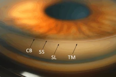
Gonioscopy is the technique for directly visualizing the anterior chamber angle, a structure that is entirely invisible on routine slit-lamp examination without special optics.
The angle sits hidden behind the limbus specifically because of total internal reflection at the corneal surface, and a goniolens is what overcomes that optical barrier and lets the examiner actually see the structures that determine how aqueous humor drains from the eye.
Skipping gonioscopy in a patient with elevated pressure or suspected angle pathology means working with an important piece of the diagnostic picture simply missing.
Despite its diagnostic importance, gonioscopy is also one of the more commonly skipped parts of a comprehensive eye exam, largely because the skill takes real practice to perform confidently and efficiently.
Why the Angle Cannot Be Seen Directly
Light reflecting off the anterior chamber angle strikes the corneal surface at an angle steep enough that it undergoes total internal reflection rather than exiting the eye toward an observer.
A goniolens changes the optical path, either through direct contact with a curved surface that eliminates this reflection or through internal mirrors in an indirect lens that redirect the angle’s image toward the examiner.
Without one of these approaches, the angle remains optically inaccessible no matter how good the slit-lamp or how favorable the lighting conditions are.
Direct Versus Indirect Gonioscopy
Direct gonioscopy uses a dome-shaped lens, most often for examination under anesthesia or intraoperative use, and gives a wide, panoramic view without needing to rotate the lens around the eye.
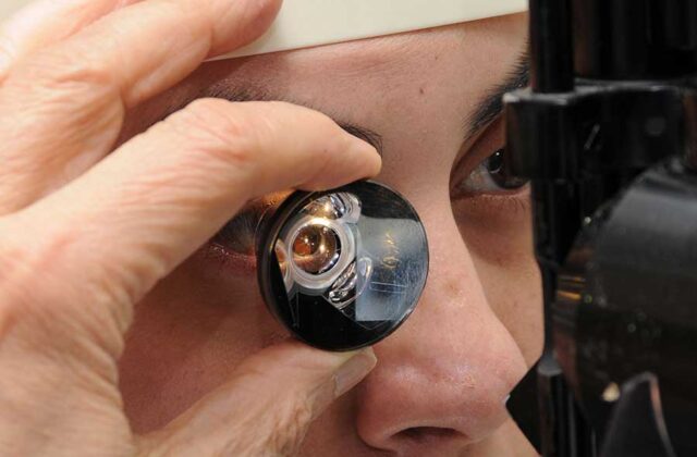
Indirect gonioscopy, the far more commonly used office technique, uses a lens with internal mirrors, most often a Goldmann or Zeiss-type lens, viewed through the slit lamp with the examiner rotating the lens or their viewing angle to inspect each quadrant in turn.
Indirect lenses vary in whether they require a coupling solution and in how much corneal indentation they allow, which matters clinically since some clinical questions specifically require the ability to indent the cornea, discussed further below.
A four-mirror lens, small enough to avoid needing coupling solution and light enough for indentation technique, has become a common office choice precisely because of that flexibility.
All-fit Slit-Lamp Adapter
Record and share exactly what you see at the slit lamp. One adapter fits any slit lamp or surgical microscope — and any smartphone.
From Choroida — the team behind this siteWhat Is Being Graded
The Shaffer and Spaeth systems are the two most widely used grading schemes, both describing the angle in terms of how open it is and which anatomic structures are visible.
The Shaffer system grades the angle from 0 to 4 based on the angular width between the iris and the trabecular meshwork, with lower grades indicating a narrower, more occludable angle.
- Grade 4: wide open, ciliary body band easily visible
- Grade 3: open, with the scleral spur visible
- Grade 2: moderately narrow, trabecular meshwork visible but the angle recess is not
- Grade 1: very narrow, only Schwalbe’s line and perhaps the top of the trabecular meshwork visible
- Grade 0: closed, iridocorneal contact with no angle structures visible
Clinical Importance
Distinguishing open-angle from angle-closure glaucoma is gonioscopy’s single most important clinical function, since the two conditions, despite sometimes producing similar pressure elevations, have entirely different mechanisms and treatments.
Indentation gonioscopy, applying gentle pressure with a smaller-diameter lens, distinguishes appositional angle closure, which opens with indentation, from synechial closure, where peripheral anterior synechiae keep the angle closed regardless of applied pressure.
Neovascularization of the angle, a sign of severe retinal ischemia in conditions like proliferative diabetic retinopathy or central retinal vein occlusion, is only reliably detected through direct gonioscopic examination, and missing it can mean missing the window to treat neovascular glaucoma before it becomes refractory.
Angle recession after blunt trauma, another finding visible only on gonioscopy, identifies eyes at long-term risk of a delayed glaucoma that can develop years after the original injury.
When to Perform It
Gonioscopy is indicated in essentially every patient being newly evaluated for glaucoma or elevated intraocular pressure, since the finding directly changes the differential diagnosis and management plan.
It should also be repeated periodically in known glaucoma patients, since angle configuration can change over time, particularly with cataract progression narrowing the angle further or after any intraocular surgery that alters anterior chamber anatomy.
Documentation matters as much as performance: recording the grade and any notable findings by quadrant, rather than a single overall impression, gives a much more useful record for comparison at future visits.


Document what you see
Two smartphone imaging tools built for everyday clinic use — one for the slit lamp, one for the fundus.
From Choroida — the team behind this siteReferences
- American Academy of Ophthalmology. Basic and Clinical Science Course, Section 10: Glaucoma.
- Shaffer RN. Primary glaucomas: gonioscopy, ophthalmoscopy, and perimetry. Transactions of the American Academy of Ophthalmology and Otolaryngology.
- Spaeth GL. The normal development of the human anterior chamber angle: a new system of descriptive grading. Transactions of the Ophthalmological Societies of the United Kingdom.
Test yourself
A few questions straight from this article.
-
Why is the anterior chamber angle invisible on routine slit-lamp examination?
Light reflecting off the angle strikes the corneal surface steeply enough to undergo total internal reflection rather than exiting toward an observer, so the angle stays optically inaccessible however good the slit lamp is. -
How does an indirect goniolens make the anterior chamber angle visible?
An indirect lens, most often a Goldmann or Zeiss-type, uses internal mirrors to redirect the angle's image toward the examiner at the slit lamp. -
What distinguishes direct gonioscopy from the usual office technique?
Direct gonioscopy uses a dome-shaped lens, most often under anesthesia or intraoperatively, giving a wide panoramic view without rotating the lens around the eye. -
In the Shaffer grading system, which finding corresponds to a grade 3 angle?
Shaffer grade 3 is an open angle with the scleral spur visible. Grade 4 shows the ciliary body band easily, grade 1 only Schwalbe's line, and grade 0 is closed with iridocorneal contact. -
Which change can narrow the angle over time, making repeat gonioscopy necessary?
Angle configuration changes over time, particularly with cataract progression narrowing the angle further, or after intraocular surgery that alters anterior chamber anatomy, so gonioscopy is repeated periodically. -
What is gonioscopy's single most important clinical function?
The two conditions can produce similar pressure elevations but have entirely different mechanisms and treatments, so distinguishing them is gonioscopy's most important role. -
In a narrow angle, how is appositional closure separated from synechial closure?
Indentation gonioscopy applies gentle pressure with a smaller-diameter lens: an appositionally closed angle opens, while peripheral anterior synechiae keep a synechially closed angle shut regardless of pressure. -
Which sign of severe retinal ischemia is reliably detected only on gonioscopy?
Angle neovascularization, a sign of severe ischemia in conditions such as proliferative diabetic retinopathy and central retinal vein occlusion, is only reliably detected gonioscopically, and missing it can mean missing the window to treat neovascular glaucoma. -
What is the long-term significance of angle recession after blunt ocular trauma?
Angle recession, visible only on gonioscopy, identifies eyes at long-term risk of a delayed glaucoma that can appear years after the original injury. -
How should gonioscopy findings be recorded for useful comparison at later visits?
Recording the grade and any notable findings quadrant by quadrant, rather than one overall impression, gives a far more useful record for comparison at future visits.

Writes and reviews the clinical articles on Educate. Every post is checked against peer-reviewed sources before it goes live.


