
Two kinds of iris nodule are described in granulomatous anterior uveitis: Koeppe nodules, at the pupil margin, and Busacca nodules, on the surface of the iris stroma.
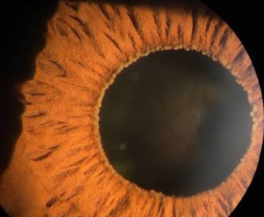
Both are collections of inflammatory cells, and both are signs that the uveitis has a granulomatous character, which narrows the differential diagnosis.
The two nodules
Koeppe nodules
These are small, translucent, gray-white nodules along the border of the pupil.
They are often arranged in a row around the pupillary margin.
They may be associated with posterior synechiae, since they sit where the iris touches the lens.
They are found in granulomatous and, to a lesser degree, non-granulomatous uveitis.
Busacca nodules
Busacca nodules lie on the anterior surface of the iris, in the stroma, within the collarette or farther peripheral.
They are larger, more opaque, and fluffier than Koeppe nodules, and they are a hallmark of granulomatous disease.
They can be the same color as the iris or paler.
What they are made of
Both consist of aggregates of lymphocytes, macrophages and epithelioid cells.
They are inflammatory nodules, and in granulomatous disease they can contain multinucleated giant cells.
They tend to resolve as the inflammation settles, and they may leave small areas of atrophy.
All-fit Slit-Lamp Adapter
Record and share exactly what you see at the slit lamp. One adapter fits any slit lamp or surgical microscope — and any smartphone.
From Choroida — the team behind this siteConditions in which they are seen
- Sarcoidosis, probably the commonest cause in many series (see sarcoid uveitis)
- Tuberculosis and syphilis
- Sympathetic ophthalmia and Vogt-Koyanagi-Harada disease (see Dalen-Fuchs nodules)
- Herpetic and other viral uveitis, in some cases
- Fungal and parasitic infections
- Lens-induced uveitis (see lens-induced uveitis)
They are often accompanied by mutton-fat keratic precipitates, posterior synechiae, and vitreous inflammation (see keratic precipitates in uveitis).
How they differ from other iris nodules
- Lisch nodules in neurofibromatosis type 1 are smooth, dome-shaped, translucent melanocytic hamartomas without inflammation (see neurofibromatosis type 1)
- Brushfield spots in Down syndrome are white-gray spots at the periphery of the iris (see Brushfield spots)
- Iris pearls are small, white, translucent nodules seen in leprosy
- Iris tumors and cysts are not associated with inflammation (see iris cyst classification)
The presence of cells in the anterior chamber and a red eye makes inflammation the likely cause.
Evaluation
- Slit-lamp examination to characterize the nodules and the presence of keratic precipitates
- Systemic work-up in granulomatous uveitis, including chest imaging, serology for syphilis, tuberculosis testing, and, depending on the history, other tests
- Fundus examination for chorioretinal lesions and vascular sheathing
Treatment
Topical corticosteroids and cycloplegics control the inflammation and relieve pain, with systemic therapy for cases with posterior involvement or an underlying systemic disease.
Nodules fade with treatment, and the treatment is directed at the underlying cause.
How to grade the nodules and the inflammation
Record the number and position of the nodules, and describe their relation to the pupil margin and the iris collarette. Grade the anterior chamber cells and flare using a standard scale, and note the size and distribution of keratic precipitates, since large, greasy "mutton-fat" precipitates and iris nodules together strongly suggest granulomatous disease. Measure the intraocular pressure, and examine the angle for nodules or synechiae.
Tests to consider
The choice of tests depends on the history. Chest imaging and serum angiotensin-converting enzyme may be requested for suspected sarcoidosis, while syphilis serology and tests for tuberculosis are appropriate in many cases of granulomatous uveitis. A careful history of travel, exposure, and systemic symptoms points the way, and a rheumatologist or an internal medicine specialist can help with the work-up.
Treatment and follow-up
Topical steroids at a frequency that matches the inflammation, cycloplegics for comfort, and a plan for a slow taper are standard. Watch the intraocular pressure carefully. Posterior synechiae can form quickly if inflammation is strong, so dilate the pupil regularly during the acute phase. Chronic granulomatous disease needs long-term monitoring, since relapses are common.
Pitfalls
- Do not assume that the presence of nodules proves sarcoidosis, because several infections and inflammatory diseases produce the same picture
- Do not forget that Koeppe nodules can occur in nongranulomatous disease
- Do not mistake iris pearls or Lisch nodules for inflammatory nodules, since they are not associated with cells in the chamber
- Do not stop treatment too early, since nodules often fade before the underlying inflammation has settled
Slit-lamp technique
Examine the iris with a narrow, oblique beam and moderate magnification, and look at the pupil margin first, then at the collarette and the periphery. Nodules are easier to see when the pupil is not fully dilated, because dilation stretches the iris and flattens them. Photographs or detailed drawings allow the nodules to be followed over time, which is a useful way to judge the response to treatment, and a change in their number often tracks changes in the anterior chamber activity.
Prognosis
Prognosis depends on the cause.
Chronic granulomatous uveitis tends to relapse, and long-term monitoring for glaucoma, cataract, and macular edema is needed.


Document what you see
Two smartphone imaging tools built for everyday clinic use — one for the slit lamp, one for the fundus.
From Choroida — the team behind this siteReferences
- Foster CS, Vitale AT. Diagnosis and Treatment of Uveitis. 2nd ed. New Delhi: Jaypee; 2013.
- Rothova A, Buitenhuis HJ, Meenken C, et al. Uveitis and systemic disease. Br J Ophthalmol. 1992;76:137-141.
- Herbort CP, Rao NA, Mochizuki M. International criteria for the diagnosis of ocular sarcoidosis. Ocul Immunol Inflamm. 2009;17:160-169.
- Jabs DA, Nussenblatt RB, Rosenbaum JT. Standardization of Uveitis Nomenclature (SUN) Working Group. Standardization of uveitis nomenclature for reporting clinical data. Am J Ophthalmol. 2005;140:509-516.
