
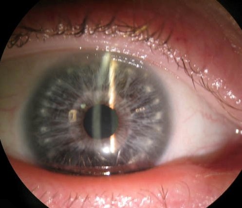
Brushfield spots are small, grayish-white or light-colored specks arranged in a ring on the peripheral iris, named after Thomas Brushfield, who first described their association with Down syndrome (trisomy 21) in 1924.
They are one of a handful of ocular findings that can prompt a clinician to consider a chromosomal diagnosis in a newborn before genetic testing results are back, though the spots themselves are harmless and require no treatment — their clinical value lies entirely in what they suggest about the patient, not in any effect they have on vision.
What They Are
Histologically, Brushfield spots represent focal areas of iris stromal hyperplasia interspersed with normal or hypoplastic stroma, creating the speckled appearance seen on exam.
They sit in the peripheral third of the iris, arranged in a roughly concentric ring, and are usually most visible in lighter-colored irides.
A very similar-appearing finding, sometimes called Kunkmann-Wolffian bodies, occurs in the general population without any chromosomal association, which is an important caveat: Brushfield spots alone are not diagnostic of Down syndrome, only suggestive.
The finding is entirely stable over a patient’s lifetime once present, neither growing nor changing character, which is part of why it functions well as a fixed, reliable clinical marker rather than something that needs to be watched for progression the way some other iris findings do.
Their number and prominence vary considerably from one affected individual to the next, and some patients with confirmed Down syndrome have too few or too subtle spots to be reliably appreciated even on careful slit-lamp exam, which is a further reason the finding functions as a supportive clue rather than a required diagnostic criterion.
Association with Down Syndrome
Brushfield spots are reported in a substantial proportion of individuals with Down syndrome, though estimates vary across studies and populations, and they are also seen — less commonly — in the general population without trisomy 21.
Their prevalence and visibility are influenced by iris pigmentation, being easier to detect in lighter irides and less so in darkly pigmented ones, which limits their usefulness as a universal screening sign.
They are one of several ocular features associated with Down syndrome, alongside upward-slanting palpebral fissures, epicanthal folds, and a higher prevalence of refractive error, strabismus, nystagmus, and keratoconus later in life.
No single feature is diagnostic on its own; the overall clinical gestalt — combined with confirmatory karyotyping — is what establishes the diagnosis.
Because pigmentation-related visibility varies so much between patients, a negative or unremarkable iris exam in a newborn with other features suggestive of Down syndrome should never be used to argue against the diagnosis; the absence of a visible spot pattern carries far less diagnostic weight than its presence.
All-fit Slit-Lamp Adapter
Record and share exactly what you see at the slit lamp. One adapter fits any slit lamp or surgical microscope — and any smartphone.
From Choroida — the team behind this siteAssociated Ocular Findings in Down Syndrome
- Refractive errors, particularly high hyperopia, at rates well above the general population
- Strabismus and nystagmus
- Keratoconus, with an earlier average age of onset and higher prevalence than in the general population
- Congenital cataract
- Blepharitis and epiphora from lid and lacrimal anomalies
Because keratoconus in particular can progress rapidly and is compounded by frequent eye rubbing (itself associated with the underlying atopic and dermatologic tendencies common in Down syndrome), regular ophthalmic follow-up is recommended for this population well beyond infancy.
Differential Diagnosis
- Kunkmann-Wolffian bodies — a similar-appearing but clinically insignificant finding in individuals without Down syndrome
- Lisch nodules — associated with neurofibromatosis type 1, typically more numerous, dome-shaped, and gelatinous-appearing rather than flat and speckled
- Iris freckles (nevi) — usually fewer, larger, and more irregularly distributed than the fine, ring-like pattern of Brushfield spots
Distinguishing Brushfield spots from Lisch nodules matters clinically, since the two point toward entirely different underlying diagnoses.
The pattern (concentric ring versus scattered) and the dome-shaped, glistening quality of Lisch nodules are usually enough to separate them on slit-lamp exam, and the broader clinical context — the facial and systemic features accompanying each condition — is usually equally telling well before any detailed iris examination is performed.
Clinical Significance
In a newborn or infant with other features suggestive of Down syndrome — hypotonia, characteristic facial features, a single palmar crease — the presence of Brushfield spots on a routine eye exam can support the clinical suspicion while formal karyotyping is pending, though they are not, on their own, sufficient to make or exclude the diagnosis.
Their real ongoing value is as a reminder to screen this population more broadly and more regularly for the ocular conditions that do threaten vision — refractive error, strabismus, and especially keratoconus — since early detection and treatment of those conditions meaningfully changes visual outcomes.
Management
Brushfield spots themselves require no treatment of any kind, since they cause no visual symptoms and carry no risk of progression or complication.
The practical management task once they are noted is entirely about the surrounding clinical picture: confirming or pursuing the underlying chromosomal diagnosis when not already established, and, once Down syndrome is confirmed, ensuring the child is enrolled in the more intensive, longitudinal ophthalmic screening schedule appropriate for this population, given the meaningfully elevated rates of refractive error, strabismus, and keratoconus described above.


Document what you see
Two smartphone imaging tools built for everyday clinic use — one for the slit lamp, one for the fundus.
From Choroida — the team behind this siteReferences
- Brushfield T. Mongolism (mongolian imbecility). Proceedings of the Royal Society of Medicine.
- Wong V, Ho D. Ocular abnormalities in Down syndrome: an analysis of 140 Chinese children. Pediatric Neurology.
- Haugen OH, Høvding G, Lundström I. Refractive development in children with Down syndrome. Acta Ophthalmologica Scandinavica.
- American Academy of Ophthalmology. Pediatric Ophthalmology and Strabismus, Basic and Clinical Science Course, Section 6.
- Kim JH, Hwang JM. Ophthalmologic manifestations in patients with Down syndrome. Korean Journal of Ophthalmology.
Test yourself
A few questions straight from this article.
-
What is the histological basis of Brushfield spots on the iris?
The speckled appearance comes from focal areas of iris stromal hyperplasia interspersed with normal or hypoplastic stroma. -
Where on the iris are Brushfield spots typically arranged?
Brushfield spots sit in the peripheral third of the iris in a roughly concentric ring, and are usually most visible in lighter-coloured irides. -
How do Brushfield spots behave over a patient's lifetime?
The finding neither grows nor changes character once present, which is why it works as a fixed clinical marker rather than something watched for progression. -
Which factor most limits detection of Brushfield spots on examination?
Visibility is influenced by iris pigmentation: the spots are easier to see in lighter irides and harder in darkly pigmented ones, which limits their value as a universal screening sign. -
What are Kunkmann-Wolffian bodies in relation to Brushfield spots?
Kunkmann-Wolffian bodies look very similar but occur in the general population with no chromosomal association, so Brushfield spots alone are suggestive rather than diagnostic. -
Which feature on slit-lamp exam favours Lisch nodules over Brushfield spots?
Lisch nodules of neurofibromatosis type 1 are typically more numerous, dome-shaped and gelatinous, and scattered, rather than the flat speckled ring of Brushfield spots. -
Which ocular condition in Down syndrome most justifies ophthalmic follow-up beyond infancy?
Keratoconus occurs earlier and more often in Down syndrome, can progress rapidly, and is compounded by frequent eye rubbing, so regular follow-up is recommended well beyond infancy. -
A newborn has features of Down syndrome but an unremarkable iris exam. How should this be interpreted?
Because visibility varies so much with pigmentation, absence of a spot pattern should never be used to argue against Down syndrome; presence carries far more diagnostic weight than absence. -
What treatment do Brushfield spots themselves require?
The spots are harmless, visually silent and carry no risk of progression or complication, so no treatment of the spots themselves is needed. -
What is the ongoing clinical value of identifying Brushfield spots in a child?
Their real value is as a reminder to screen this population regularly for refractive error, strabismus and especially keratoconus, where early treatment changes visual outcomes.
