
Neurofibromatosis type 1 is an autosomal dominant disorder of the NF1 tumour suppressor gene, and the eye is involved often enough, and distinctively enough, that ophthalmic findings are written directly into the diagnostic criteria rather than being an incidental association.
Lisch nodules, in particular, are close to pathognomonic once seen clearly at the slit lamp, which makes ophthalmic examination a genuinely useful contributor to diagnosing a systemic disease, not just a search for ocular complications of a diagnosis already made elsewhere.
Diagnostic Criteria
Diagnosis requires two or more of a defined set of features, several of which are ophthalmic or periocular: six or more café-au-lait macules, two or more neurofibromas of any type or one plexiform neurofibroma, freckling in the axillary or inguinal region, two or more Lisch nodules, a distinctive osseous lesion, an optic pathway glioma, and a first-degree relative with confirmed NF1.
Lisch nodules and optic pathway glioma are the two specifically ophthalmic criteria, which is a large part of why a careful eye examination, including slit-lamp assessment of the iris, is a routine part of the diagnostic workup in a child with suspected NF1.
Ocular Manifestations
Lisch Nodules
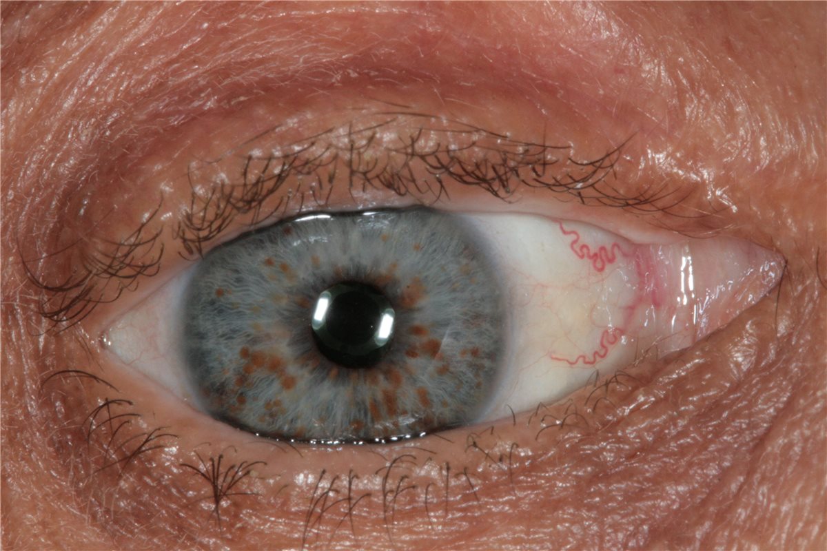
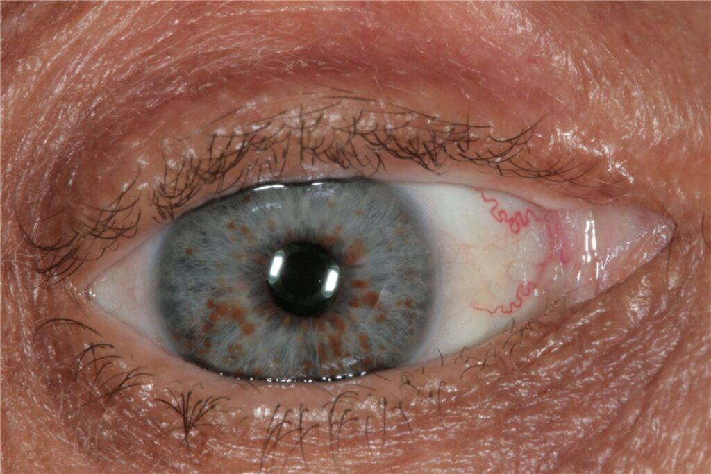
These are small, smooth, dome-shaped melanocytic hamartomas scattered across the iris surface, increasing in number with age, and present in the great majority of adults with NF1 by adulthood, even though they may be absent or sparse in early childhood.
They cause no visual symptoms and require no treatment themselves; their value is purely diagnostic, as a specific and easily examined marker of the underlying condition.
Optic Pathway Glioma
A low-grade pilocytic astrocytoma of the optic nerve, chiasm, or optic radiations occurs in a meaningful minority of children with NF1, most commonly identified in the first decade of life.
Many optic pathway gliomas in NF1 remain stable or grow very slowly and never cause symptoms, which is a genuinely different biology from sporadic optic gliomas and is central to how surveillance and treatment decisions are made in this specific population.
Plexiform Neurofibroma of the Eyelid
A diffuse, infiltrative neurofibroma can involve the eyelid, classically producing a characteristic S-shaped deformity of the upper lid margin along with ptosis, and can be associated with ipsilateral orbital or facial bony changes, including sphenoid wing dysplasia.
Other Findings
Choroidal abnormalities, detectable on infrared or near-infrared imaging as bright patchy nodules, are increasingly recognised and used as an additional supportive diagnostic feature, though they are not yet part of the formal diagnostic criteria. Congenital glaucoma can occur, particularly associated with ipsilateral eyelid plexiform neurofibroma.
All-fit Slit-Lamp Adapter
Record and share exactly what you see at the slit lamp. One adapter fits any slit lamp or surgical microscope — and any smartphone.
From Choroida — the team behind this siteScreening Approach
Annual ophthalmic examination through early childhood is recommended for children with NF1, given the age-related emergence of both Lisch nodules and the peak incidence window for optic pathway glioma.
Examination should specifically include visual acuity, colour vision, pupillary assessment, and dilated fundus examination looking for optic disc pallor or swelling, alongside slit-lamp assessment of the iris.
MRI of the optic pathways is used to investigate any concerning visual findings and is considered for baseline or surveillance imaging in some centres, guided by local protocols and the clinical picture rather than performed routinely and unconditionally in every child.
Differential Diagnosis
- Neurofibromatosis type 2, which has essentially no Lisch nodules but instead carries a strong association with bilateral vestibular schwannoma and posterior subcapsular cataract, a very different ocular profile from NF1
- Sporadic optic glioma without NF1, generally more aggressive and requiring closer monitoring and more active treatment consideration than the typically indolent NF1-associated form
- Iris naevi or freckles, which can superficially resemble Lisch nodules but lack their characteristic smooth, dome-shaped, translucent appearance on slit-lamp examination
- McCune-Albright syndrome, which can share café-au-lait pigmentation but has a different pattern of skin lesion borders and an entirely different set of associated endocrine findings
Management
Lisch nodules require no treatment. Optic pathway glioma is managed with observation in stable, asymptomatic cases, reserving chemotherapy for tumours showing radiological progression or causing visual decline, since the generally indolent behaviour of NF1-associated gliomas often does not warrant the risks of upfront treatment.
Plexiform neurofibroma of the eyelid is managed surgically when it causes visually significant ptosis or amblyopia, though complete resection is often difficult given the diffuse, infiltrative nature of these lesions, and recurrence after debulking surgery is common.
Amblyopia should be screened for and treated in any child with ptosis or astigmatism from eyelid or orbital involvement, applying standard occlusion therapy principles alongside management of the underlying structural problem.
Prognosis
Vision is preserved in most patients with NF1, since Lisch nodules themselves are entirely benign and most optic pathway gliomas remain stable without treatment.
Visual outcome in the minority with progressive optic pathway glioma depends on how promptly progression is identified through surveillance and treated, which is the main rationale for structured ophthalmic follow-up through childhood rather than examination only when symptoms appear.
Eyelid plexiform neurofibroma can be more functionally significant if it causes amblyopia from ptosis or induced astigmatism, making early recognition and, where indicated, amblyopia treatment more consequential for long-term vision than the neurofibroma itself.


Document what you see
Two smartphone imaging tools built for everyday clinic use — one for the slit lamp, one for the fundus.
From Choroida — the team behind this siteReferences
- Legius E, Messiaen L, Wolkenstein P, et al. Revised diagnostic criteria for neurofibromatosis type 1 and Legius syndrome. Genetics in Medicine. 2021.
- Lewis RA, Gerson LP, Axelson KA, et al. von Recklinghausen neurofibromatosis. II. Incidence of optic gliomata. Ophthalmology. 1984.
- Listernick R, Ferner RE, Liu GT, Gutmann DH. Optic pathway gliomas in neurofibromatosis-1: controversies and recommendations. Annals of Neurology. 2007.
- Neurofibromatosis Type 1. EyeWiki, American Academy of Ophthalmology.
- Neurofibromatosis Type 1. StatPearls, NCBI Bookshelf.
Test yourself
A few questions straight from this article.
-
What is the inheritance pattern of neurofibromatosis type 1?
Neurofibromatosis type 1 is an autosomal dominant disorder of the NF1 tumour suppressor gene. -
Which two diagnostic criteria for neurofibromatosis type 1 are specifically ophthalmic?
Lisch nodules and optic pathway glioma are the two ophthalmic entries in the diagnostic criteria, which is why slit-lamp iris assessment belongs in the workup. -
How many cafe-au-lait macules are required as a diagnostic criterion in neurofibromatosis type 1?
Six or more cafe-au-lait macules count as one criterion; diagnosis requires two or more criteria from the defined set. -
What are Lisch nodules in neurofibromatosis type 1?
They are small, smooth, dome-shaped melanocytic hamartomas scattered over the iris that increase in number with age, cause no visual symptoms and need no treatment. -
Which tumour type is the optic pathway glioma of neurofibromatosis type 1?
It is a low-grade pilocytic astrocytoma of the optic nerve, chiasm or radiations, most often identified in the first decade of life. -
Which eyelid sign suggests plexiform neurofibroma in neurofibromatosis type 1?
A diffuse infiltrative eyelid neurofibroma classically gives an S-shaped upper lid margin with ptosis, and may accompany ipsilateral bony changes such as sphenoid wing dysplasia. -
Which ocular finding characterises neurofibromatosis type 2 rather than type 1?
Neurofibromatosis type 2 has essentially no Lisch nodules but is strongly associated with posterior subcapsular cataract and bilateral vestibular schwannoma. -
How do the choroidal abnormalities of neurofibromatosis type 1 appear on near-infrared imaging?
Choroidal abnormalities appear as bright patchy nodules on infrared or near-infrared imaging and are used as supportive evidence, though not yet a formal diagnostic criterion. -
How is a stable, asymptomatic optic pathway glioma in neurofibromatosis type 1 managed?
These tumours are typically indolent, so stable asymptomatic cases are observed and chemotherapy is reserved for radiological progression or visual decline. -
What ophthalmic follow-up is recommended for children with neurofibromatosis type 1?
Annual examination through early childhood covers both the age-related emergence of Lisch nodules and the peak window for optic pathway glioma.

Writes and reviews the clinical articles on Educate. Every post is checked against peer-reviewed sources before it goes live.


