
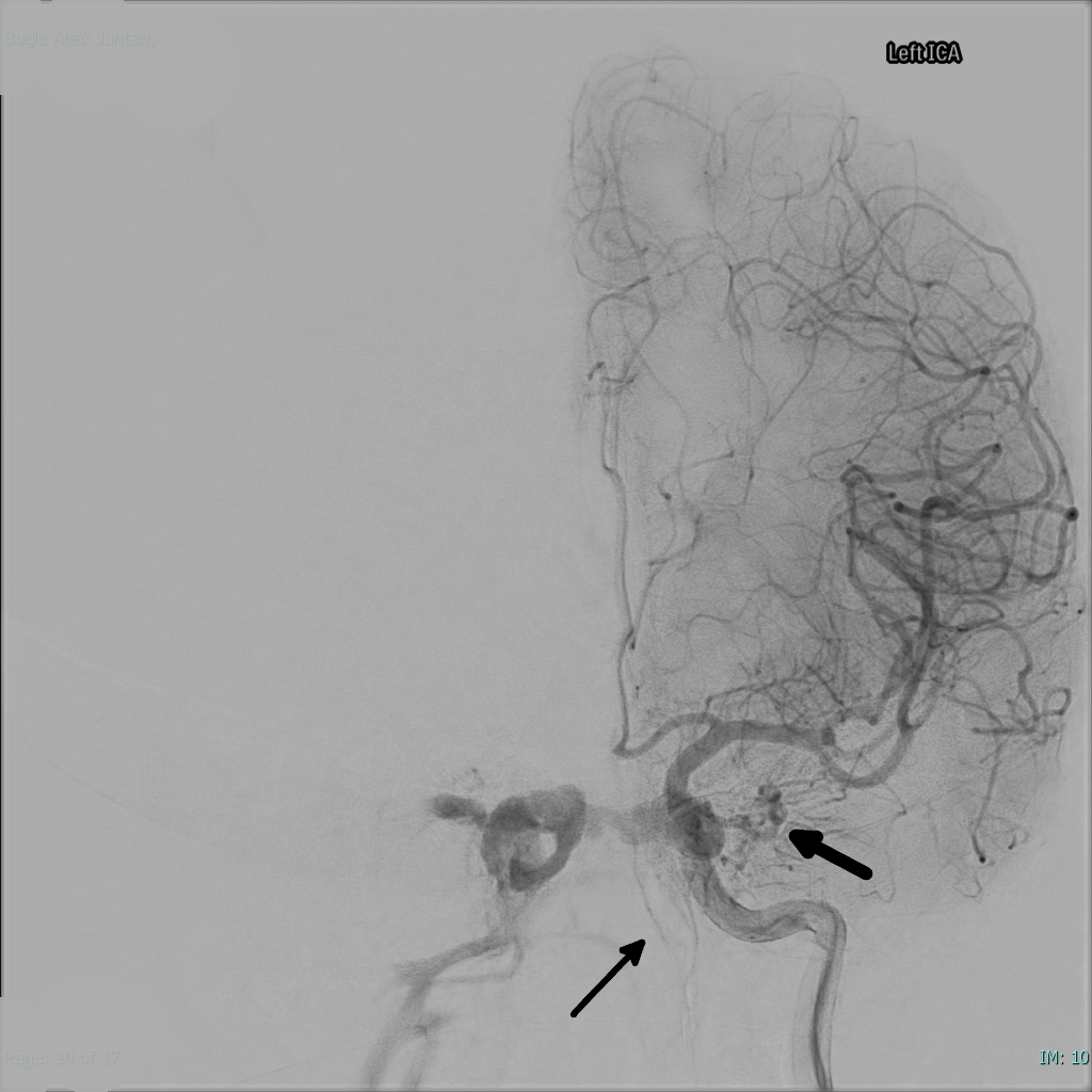
A carotid-cavernous fistula (CCF) is an abnormal connection between the carotid arterial system and the cavernous sinus, allowing high-pressure arterial blood to flow directly into a venous structure that was never built to carry it.
The ocular findings that follow, pulsatile proptosis, chemosis, and an orbital bruit, are really downstream signs of venous congestion. Recognising them for what they are is what gets a patient to the right angiography suite instead of a slower workup for orbital inflammation.
Fistulas are classified by their anatomy and flow rate, and this classification drives almost everything about presentation, urgency, and treatment.
Classification
Direct fistulas involve a direct communication between the internal carotid artery itself and the cavernous sinus, most often following head trauma or rupture of an intracavernous carotid aneurysm. Flow through a direct fistula is high, and the onset of symptoms is typically abrupt.
Indirect, or dural, fistulas instead involve communication between meningeal branches of the internal or external carotid artery and the cavernous sinus. These are lower-flow and tend to arise spontaneously, more often in older women, sometimes in the setting of connective tissue disease.
The Barrow classification refines this further into four types (A through D) based on the specific arterial feeders involved, which matters mainly for endovascular planning rather than for the initial clinical assessment.
Pathophysiology
Once a fistula forms, arterial pressure is transmitted directly into the cavernous sinus, and from there retrograde into the superior and inferior ophthalmic veins. This raises episcleral venous pressure, which is responsible for most of the signs seen at the slit lamp: dilated, tortuous, corkscrew episcleral vessels, conjunctival chemosis, and often a genuinely elevated intraocular pressure from impaired aqueous outflow.
Orbital venous congestion produces proptosis, and because the sinus receives pulsatile arterial inflow, that proptosis is itself pulsatile, sometimes visibly so, sometimes only detectable on palpation or with a stethoscope over the closed lid.
Cranial nerves III, IV, V1, V2, and VI all pass through or near the cavernous sinus, so any combination of ophthalmoplegia and facial sensory change can accompany the orbital findings, depending on which nerves are compressed or stretched by the congested sinus. Sixth nerve involvement is particularly common given the abducens nerve’s course directly through the sinus rather than along its lateral wall.
All-fit Slit-Lamp Adapter
Record and share exactly what you see at the slit lamp. One adapter fits any slit lamp or surgical microscope — and any smartphone.
From Choroida — the team behind this siteClinical Presentation
Direct, high-flow fistulas tend to present acutely and dramatically: sudden proptosis, redness, chemosis, and diplopia, often following clear head trauma, which makes the diagnosis relatively easy to suspect.
Indirect fistulas are the more diagnostically challenging group. Onset is gradual, chemosis and injection can look very much like chronic conjunctivitis or thyroid eye disease, and a patient may be treated for one of those conditions for weeks before anyone auscultates the orbit or asks about a bruit.
On examination, look specifically for corkscrew episcleral vessels, which are a genuinely distinctive sign once you have seen them; a pulsatile proptosis that may reduce with gentle globe compression; chemosis, sometimes severe enough to prolapse over the lid margins; and elevated intraocular pressure from raised episcleral venous pressure.
An orbital or cranial bruit, audible with a stethoscope over the closed eye or temple, is present in many but not all cases and is worth listening for specifically rather than assuming its absence rules out the diagnosis. Diplopia from cranial nerve involvement, most often the sixth nerve, is common.
A dilated fundus examination can show venous congestion, disc swelling, or retinal haemorrhages in more severe or longstanding cases.

Diagnostic Evaluation
CT or MRI of the orbit and brain typically shows an enlarged superior ophthalmic vein and proptosis, and can suggest cavernous sinus enlargement, though cross-sectional imaging alone does not always distinguish a fistula confidently from other causes of orbital congestion.
Doppler ultrasound of the orbit can demonstrate arterialised, reversed flow in the superior ophthalmic vein, which is a useful and non-invasive supporting finding.
Cerebral catheter angiography remains the definitive investigation. It confirms the diagnosis, defines the exact fistula anatomy and feeding vessels, and is usually performed as the first step of a combined diagnostic-and-treatment procedure, since most fistulas requiring intervention are treated endovascularly at the same sitting.
Differential Diagnosis
- Thyroid eye disease, which can mimic chronic proptosis and chemosis but lacks pulsatility, corkscrew vessels, or a bruit
- Orbital cellulitis or idiopathic orbital inflammation, generally more acutely painful and without the vascular signs described above
- Cavernous sinus thrombosis, which shares some of the cranial nerve findings but follows a different, typically more septic or acutely painful, clinical course
- Orbital venous or lymphatic malformation, which can also present with proptosis but again without pulsatility or a bruit
Pulsatility, a bruit, and corkscrew episcleral vessels together are unusual enough that when all three are present, the differential narrows quickly to a fistula.
Management
Some low-flow indirect fistulas resolve spontaneously or with conservative management, and observation is reasonable in mild cases without threatened vision, uncontrolled glaucoma, or significant diplopia. This is not the default approach for every patient, but it is a legitimate option in carefully selected, non-urgent presentations.
Endovascular embolisation, most often via a transvenous approach through the inferior petrosal sinus, is now the primary treatment for most fistulas requiring intervention, aiming to occlude the abnormal connection while preserving normal arterial flow. Direct, high-flow fistulas usually need more urgent treatment given their rate of visual and neurological complications.
Surgical or radiosurgical approaches are reserved for the minority of cases not amenable to an endovascular approach.
Elevated intraocular pressure is managed medically in the interim with standard glaucoma therapy, recognising that definitive control of pressure usually depends on closing the fistula itself rather than on drops alone. Indications for more urgent intervention include progressive visual loss, uncontrolled intraocular pressure, and rapidly worsening proptosis or chemosis threatening the ocular surface.
Prognosis
Most fistulas treated endovascularly achieve good closure rates with resolution or substantial improvement of the ocular signs.
Outcome depends heavily on how much secondary damage has already occurred by the time of treatment, particularly glaucomatous optic nerve damage from prolonged elevated pressure and any ischaemic retinal or optic nerve injury from chronic venous congestion.
Diplopia from cranial nerve involvement often improves once the fistula is closed, though recovery can be incomplete if compression has been longstanding.
Delayed diagnosis, especially in indirect fistulas misattributed for weeks to more common causes of red eye, is the main factor separating a good outcome from a permanent one, which is really the argument for keeping this diagnosis on the differential whenever proptosis and chemosis do not fit a more common pattern.


Document what you see
Two smartphone imaging tools built for everyday clinic use — one for the slit lamp, one for the fundus.
From Choroida — the team behind this siteReferences
- Barrow DL, Spector RH, Braun IF, et al. Classification and treatment of spontaneous carotid-cavernous sinus fistulas. Journal of Neurosurgery. 1985.
- Ellis JA, Goldstein H, Connolly ES, Meyers PM. Carotid-cavernous fistulas. Neurosurgical Focus. 2012.
- Miller NR. Diagnosis and management of dural carotid-cavernous sinus fistulas. Neurosurgical Focus. 2007.
- Carotid-Cavernous Fistula. EyeWiki, American Academy of Ophthalmology.
- Carotid Cavernous Fistula. StatPearls, NCBI Bookshelf.
Test yourself
A few questions straight from this article.
-
A carotid-cavernous fistula is an abnormal connection between which two structures?
The fistula lets high-pressure arterial blood flow directly into a venous structure never built to carry it. The ocular findings that follow are downstream signs of that venous congestion. -
Which scenario most often precedes a direct, high-flow carotid-cavernous fistula?
Direct fistulas connect the internal carotid artery itself to the cavernous sinus, usually after head trauma or rupture of an intracavernous aneurysm. Flow is high and symptom onset is typically abrupt. -
Which description fits an indirect, or dural, carotid-cavernous fistula?
Indirect fistulas connect meningeal branches of the internal or external carotid artery to the cavernous sinus. They are lower-flow, arise spontaneously more often in older women, and sometimes accompany connective tissue disease. -
Why is intraocular pressure often elevated in a carotid-cavernous fistula?
Arterial pressure passes retrograde into the superior and inferior ophthalmic veins, raising episcleral venous pressure and impairing aqueous outflow. The same mechanism produces the chemosis and dilated episcleral vessels. -
Which slit-lamp finding is most distinctive of a carotid-cavernous fistula?
Corkscrew episcleral vessels are a genuinely distinctive sign once seen, reflecting raised episcleral venous pressure. They appear alongside chemosis and a pulsatile proptosis that may reduce with gentle globe compression. -
Why is sixth nerve involvement particularly common in carotid-cavernous fistula?
The abducens nerve courses through the sinus itself, while the other nerves travel in its lateral wall. That position leaves it most exposed to compression or stretching by the congested sinus. -
Which features separate a carotid-cavernous fistula from thyroid eye disease at the bedside?
Thyroid eye disease can mimic chronic proptosis and chemosis but lacks pulsatility, corkscrew vessels and a bruit. When all three vascular signs are present the differential narrows quickly to a fistula. -
Which investigation is definitive for confirming and characterising a carotid-cavernous fistula?
Catheter angiography confirms the diagnosis and defines the exact fistula anatomy and feeding vessels. It is usually the first step of a combined diagnostic-and-treatment sitting, since most fistulas are then treated endovascularly. -
What is now the primary treatment for most carotid-cavernous fistulas requiring intervention?
Embolisation, most often transvenously through the inferior petrosal sinus, occludes the abnormal connection while preserving normal arterial flow. Surgical and radiosurgical approaches are reserved for cases unsuitable for endovascular treatment. -
Which factor most separates a good outcome from permanent damage in carotid-cavernous fistula?
Indirect fistulas are often misattributed for weeks to commoner causes of red eye. Outcome depends heavily on secondary damage already accrued, particularly glaucomatous optic nerve injury from prolonged raised pressure.

Writes and reviews the clinical articles on Educate. Every post is checked against peer-reviewed sources before it goes live.


