
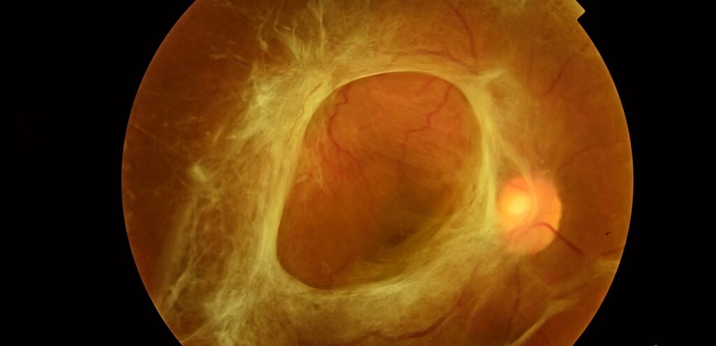
Tractional retinal detachment is the separation of the neurosensory retina from the underlying retinal pigment epithelium caused by mechanical pulling from fibrovascular or fibrous membranes on the retinal surface, rather than by fluid passing through a retinal break as in rhegmatogenous detachment.
Understanding this distinction is not just academic: the two types of detachment behave differently, progress at different rates, and are managed with fundamentally different surgical strategies, making correct identification at the outset essential to planning appropriate treatment.
Mechanism
Tractional membranes form on the retinal surface (or, in proliferative vitreoretinopathy, also beneath it) as a response to an underlying disease process, typically one involving retinal ischemia and subsequent neovascularization that provides scaffolding for fibrous tissue to organize along.
As these membranes contract over time, they exert progressive traction on the retina at their points of attachment, gradually pulling it away from the underlying RPE without any fluid entering through a break, at least in the pure, uncomplicated form of the condition.
Because this process depends on the membrane physically contracting rather than fluid actively flowing in, tractional detachment characteristically progresses far more slowly than a typical rhegmatogenous detachment, often over weeks to months rather than hours to days, which shapes both how it presents and how urgently it typically needs to be addressed.
Underlying Causes
- Proliferative diabetic retinopathy — the most common cause overall, from fibrovascular membranes growing along the posterior hyaloid face in response to retinal ischemia
- Retinopathy of prematurity, advanced stages — fibrovascular proliferation at the vascular-avascular junction
- Proliferative vitreoretinopathy — fibrocellular membrane formation, classically as a complication of prior rhegmatogenous retinal detachment or its repair
- Penetrating ocular trauma with vitreous hemorrhage and subsequent fibrous organization
- Sickle cell retinopathy, familial exudative vitreoretinopathy, and other less common proliferative retinal conditions
Fundus Explorer Pro
Photograph the retinal findings described here with the phone already in your pocket — 22 D optics and built-in illumination in one handheld unit.
From Choroida — the team behind this siteClinical Presentation
Unlike rhegmatogenous detachment, which classically presents with sudden flashes, floaters, and a curtain-like visual field defect, tractional detachment tends to progress more slowly and can be asymptomatic until it involves the macula, in a patient with pre-existing visual impairment from the underlying disease (diabetic retinopathy, for instance) who may not immediately notice further gradual decline.
This slower, quieter presentation is part of why tractional detachment is more often discovered on a scheduled retinal exam than through an urgent same-day presentation, though this is not universally true and depends on the extent and location of the traction.
This is precisely why regular, scheduled dilated retinal screening is so important in patients with proliferative diabetic retinopathy and other high-risk conditions, since a slowly progressing tractional detachment can advance a meaningful distance toward the macula between visits without producing any symptom that would otherwise prompt the patient to seek care sooner.
Exam Findings
- A concave, relatively immobile retinal surface, in contrast to the convex, more mobile appearance typical of rhegmatogenous detachment
- The detachment is usually confined to the area under traction and does not typically extend to the ora serrata, another distinguishing feature from rhegmatogenous detachment
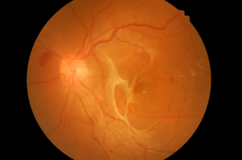
- Visible fibrovascular or fibrous membranes on the retinal surface, sometimes with associated vitreous hemorrhage obscuring the view
- Signs of the underlying disease — diabetic changes, ROP-related vascular findings, or evidence of prior trauma or surgery
Combined Tractional-Rhegmatogenous Detachment
A tractional membrane can eventually create enough force to actually tear the retina at its point of attachment, converting a pure tractional detachment into a combined tractional-rhegmatogenous detachment.
This behaves more aggressively and progresses faster than either mechanism alone.
Recognizing this combined pattern matters because it usually shifts the urgency of surgical planning, since the fluid-driven component of a rhegmatogenous detachment can extend and progress much faster than pure traction alone would, effectively converting a case that could previously be monitored into one that requires prompt surgical repair.
Differential Diagnosis
- Rhegmatogenous retinal detachment — convex, mobile, extends to the ora, associated with a visible break
- Exudative retinal detachment — smooth, shifting fluid that moves with gravity and changes shape with head position, without traction or a break, from an underlying inflammatory, neoplastic, or vascular exudative process
- Combined tractional-rhegmatogenous detachment, as described above, which shares features of both pure forms
Management
Not every tractional detachment requires immediate surgery.
A stable, peripheral detachment not threatening or involving the macula can sometimes be observed with close monitoring, in a patient with significant surgical risk from other factors.
Detachment threatening or involving the macula, or one showing clear progression on serial exams, generally requires pars plana vitrectomy with careful membrane dissection (segmentation and delamination techniques) to relieve the traction, often combined with panretinal photocoagulation for the underlying ischemic drive and, when a combined rhegmatogenous component is present, the additional standard measures used for any retinal break (endolaser, tamponade).
Surgery for tractional detachment is technically demanding, since the membranes are often adherent to a thin, already compromised retina, and the risk of iatrogenic retinal breaks during membrane removal is a genuine intraoperative concern that shapes surgical technique throughout the case.
Because the underlying disease process (diabetic retinopathy, ROP, or another proliferative condition) that produced the traction in the first place remains active after surgery unless independently controlled, ongoing treatment of that underlying condition and close postoperative surveillance for recurrent traction or new membrane formation are essential parts of long-term management, not simply a one-time surgical fix.


Document what you see
Two smartphone imaging tools built for everyday clinic use — one for the slit lamp, one for the fundus.
From Choroida — the team behind this siteReferences
- Charles S, Calzada J, Wood B. Vitreous Microsurgery.
- Machemer R, Buettner H, Norton EW, Parel JM. Vitrectomy: a pars plana approach. Transactions of the American Academy of Ophthalmology and Otolaryngology.
- American Academy of Ophthalmology. Basic and Clinical Science Course, Section 12: Retina and Vitreous.
Test yourself
A few questions straight from this article.
-
What causes the retinal separation in tractional retinal detachment?
Fibrovascular or fibrous membranes contract and pull the neurosensory retina off the pigment epithelium, without fluid entering through a break as in rhegmatogenous detachment. -
How do the membranes of tractional retinal detachment form?
Retinal ischemia provokes neovascularization, which provides scaffolding for fibrous tissue to organize along; as those membranes contract they pull on their points of attachment. -
How quickly does a pure tractional retinal detachment usually progress?
Progression depends on slow membrane contraction rather than active fluid flow, so it advances over weeks to months rather than the hours to days of a rhegmatogenous detachment. -
Which condition is the most common cause of tractional retinal detachment?
Fibrovascular membranes growing along the posterior hyaloid face in response to ischemia make proliferative diabetic retinopathy the commonest cause overall. -
What is the characteristic surface appearance of a tractional retinal detachment?
The retina is held taut by contracting membranes, giving a concave, immobile surface, unlike the convex and mobile appearance of a rhegmatogenous detachment. -
Why is tractional retinal detachment often found on a scheduled retinal examination?
Slow progression in an eye already visually impaired by the underlying disease means gradual decline can go unnoticed, which is why regular dilated screening matters in high-risk patients. -
How does an exudative retinal detachment differ from a tractional one?
Exudative detachment shows smooth, shifting subretinal fluid that moves with head position, with no traction and no retinal break. -
Why does a combined tractional-rhegmatogenous detachment change management?
When traction tears the retina at an attachment point, the added rhegmatogenous component extends far faster, turning a case that could be monitored into one needing prompt repair. -
Which tractional retinal detachment can reasonably be observed with close monitoring?
Stable peripheral traction may be watched, particularly when surgical risk is high; macula-threatening or clearly progressing detachment needs vitrectomy with segmentation and delamination. -
Why is continued follow-up essential after vitrectomy for tractional retinal detachment?
Surgery relieves the traction but does not treat the diabetic retinopathy or other proliferative process, so recurrent traction and new membranes remain possible without ongoing control.

Writes and reviews the clinical articles on Educate. Every post is checked against peer-reviewed sources before it goes live.


