
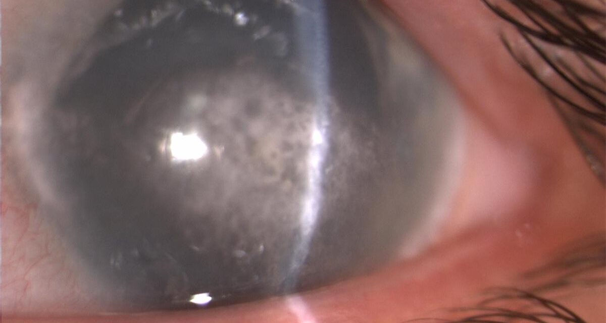
A cloudy band creeping across the front of the eye is rarely the primary disease — it is usually a scar left by something else.
Band keratopathy is calcium deposition in a horizontal band across the cornea, and it is almost always a marker of chronic ocular or systemic disease.
It develops slowly, often over months to years, and is easy to miss until it has already encroached on the visual axis.
Because it is a secondary process rather than a primary corneal disorder, effective management starts with identifying what drove the calcium there in the first place.
Treating the band alone without addressing the underlying trigger frequently leads to recurrence.
Recognizing the classic appearance — and knowing when to look for chronic uveitis, glaucoma therapy, or a systemic calcium disorder behind it — is central to managing it well.
What Is Band Keratopathy?
Band keratopathy is the deposition of calcium salts, principally calcium phosphate, within the superficial cornea — primarily in Bowman’s layer.
Its defining features include:
- A horizontal, gray-white band crossing the cornea within the interpalpebral fissure
- Characteristic round, clear holes within the band, giving a “Swiss cheese” appearance corresponding to nerve openings through Bowman’s layer
- A clear zone typically separating the band from the limbus
- Slowly progressive course, often asymptomatic until advanced
The interpalpebral distribution is a key diagnostic clue and relates directly to how the deposits form.
Epidemiology
Band keratopathy is uncommon as a primary presenting complaint but is regularly seen as a late finding in chronic ocular disease.
- Large case series report roughly a third of cases as idiopathic, with no clear identifiable trigger
- Chronic corneal edema and long-term topical glaucoma medication use are among the most frequently identified associated conditions
- An underlying systemic comorbidity is identifiable in roughly a third of cases
- Prevalence increases with the duration of any chronic ocular surface or intraocular disease
Because it develops gradually, band keratopathy is often first noticed incidentally during routine follow-up of a known chronic eye condition.
All-fit Slit-Lamp Adapter
Record and share exactly what you see at the slit lamp. One adapter fits any slit lamp or surgical microscope — and any smartphone.
From Choroida — the team behind this sitePathophysiology
The interpalpebral distribution of band keratopathy is not coincidental — it reflects the local environment of that zone.
- The exposed interpalpebral cornea undergoes more tear film evaporation than the protected, lid-covered cornea
- Evaporation locally raises tear film pH and carbon dioxide loss in this zone
- The resulting alkaline shift favors precipitation of calcium and phosphate ions from the tears and aqueous
- Calcium phosphate crystals deposit within Bowman’s layer and the superficial stroma
- Corneal nerves passing through Bowman’s layer create the characteristic clear holes within the band
Any condition that raises local or systemic calcium/phosphate levels, or that chronically disrupts the ocular surface, can accelerate this same process.
Risk Factors
Ocular Risk Factors
- Chronic anterior uveitis, particularly juvenile idiopathic arthritis-associated uveitis in children
- Chronic corneal edema from any cause
- Long-term use of topical medications containing phosphate-buffered preservatives, especially in eyes with an already compromised epithelium
- Phthisis bulbi and other chronically inflamed, blind eyes
- Prior silicone oil tamponade in eyes that have undergone vitreoretinal surgery
Systemic Risk Factors
- Hypercalcemia from any cause, including hyperparathyroidism and sarcoidosis
- Chronic renal failure with secondary hyperparathyroidism and altered calcium-phosphate metabolism
- Vitamin D toxicity
- Rare hereditary forms occurring without an identifiable ocular or systemic trigger
A meaningful minority of cases remain idiopathic even after a thorough workup, so band keratopathy alone does not always demand an exhaustive systemic investigation.
Clinical Presentation
Symptoms
- Often asymptomatic in early stages, found incidentally on slit-lamp exam
- Foreign-body sensation or irritation if the surface becomes irregular or the epithelium breaks down over calcium deposits
- Gradually decreasing vision as the band encroaches on the visual axis
- Cosmetic concern from visible corneal clouding in advanced cases
Examination Findings
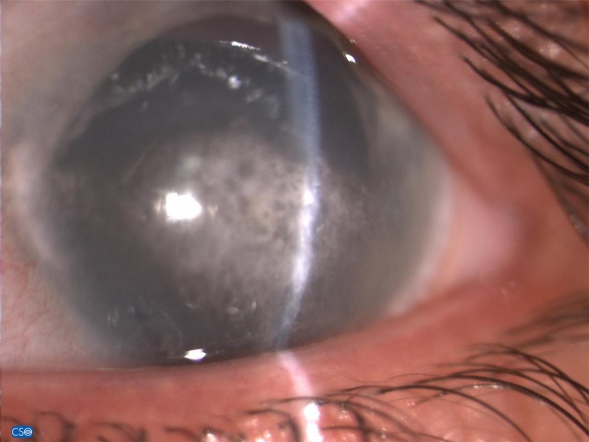
- A horizontal, chalky gray-white band spanning the interpalpebral cornea
- Small, round, clear lucencies scattered within the band — the classic Swiss-cheese pattern
- A clear gap typically visible between the band and the limbus
- Progressive encroachment toward the visual axis in longstanding or advanced cases
- Signs of the underlying cause — active or quiet uveitis, corneal edema, or a phthisical eye — should always be sought alongside the band itself
The interpalpebral location and Swiss-cheese lucencies are distinctive enough that band keratopathy is rarely mistaken for another corneal opacity once specifically looked for.
Diagnostic Evaluation
Slit-Lamp Examination
- Confirms the characteristic appearance and documents the extent of visual axis involvement
- Should always include a search for active intraocular inflammation or corneal edema as a potential driver
Systemic Workup
- Serum calcium, phosphate, and, when indicated, parathyroid hormone levels, particularly in bilateral or atypical cases without an obvious ocular cause
- Renal function testing when chronic kidney disease is suspected as a contributing factor
- A directed workup is more appropriate than a reflexive one — the extent of testing should be guided by whether an ocular cause is already evident
Identifying and treating the underlying trigger is at least as important diagnostically as characterizing the band itself.
Differential Diagnosis
Conditions that can be confused with band keratopathy include:
- Corneal arcus — a peripheral, limbus-adjacent ring rather than an interpalpebral band, and unrelated to calcium deposition
- Salzmann nodular degeneration — discrete elevated nodules rather than a flat calcific band
- Corneal scarring from prior keratitis — typically lacks the characteristic Swiss-cheese lucencies
- Corneal dystrophies with stromal deposits — usually bilateral, symmetric, and without an identifiable secondary trigger
The interpalpebral distribution combined with the round clear holes within the band is usually sufficient to distinguish band keratopathy from these mimics on slit-lamp exam alone.
Management
Treating the Underlying Cause
- Controlling active uveitis or corneal edema reduces the ongoing drive for further calcium deposition
- Correcting hypercalcemia or an underlying systemic calcium-phosphate disorder, in coordination with the treating physician
- Switching or minimizing phosphate-containing topical medications when they are a plausible contributor
EDTA Chelation
- Topical EDTA (ethylenediaminetetraacetic acid) chelation is the standard first-line treatment for visually significant band keratopathy
- Performed after removing the corneal epithelium overlying the band, with repeated EDTA application to dissolve the calcium deposits
- Effective and low-cost, with visual improvement in most treated eyes and a comparatively low need for retreatment in published series
Other Procedural Options
- Superficial keratectomy, with or without amniotic membrane transplantation, for thicker or more resistant deposits
- Phototherapeutic keratectomy (PTK) as an alternative or adjunct, though at higher cost and with a risk of inducing refractive change
Whichever technique is chosen, treatment addresses a visible consequence of chronic disease — it does not replace management of the underlying condition that caused it.
Prognosis
Visual and cosmetic outcomes after treatment are generally favorable when the underlying cause is also controlled.
- Most eyes show meaningful visual improvement after EDTA chelation or superficial keratectomy
- Recurrence is more likely when the driving ocular or systemic condition remains active or poorly controlled
- In severely damaged or phthisical eyes, band keratopathy is often a marker of poor overall visual prognosis independent of the corneal finding itself
Long-term outcome tracks the underlying disease far more than it tracks the corneal procedure chosen to clear the band.
Would you like to document anterior segment findings with your smartphone?


Document what you see
Two smartphone imaging tools built for everyday clinic use — one for the slit lamp, one for the fundus.
From Choroida — the team behind this siteReferences
- Najjar DM, Cohen EJ, Rapuano CJ, Laibson PR. EDTA chelation for calcific band keratopathy: results and long-term follow-up. American Journal of Ophthalmology. 2004.
- Al-Hity A, Ramaesh K, Lockington D. EDTA chelation for symptomatic band keratopathy: results and recurrence. Eye. 2018.
- Clinical characteristics and outcomes of band keratopathy: a 10-year retrospective analysis. Medical Hypothesis, Discovery & Innovation in Optometry. 2023.
- Jhanji V, Rapuano CJ, Vajpayee RB. Corneal calcific band keratopathy. Current Opinion in Ophthalmology. 2011.
- Band Keratopathy. EyeWiki, American Academy of Ophthalmology.
Test yourself
A few questions straight from this article.
-
In band keratopathy, which material is deposited and in which corneal layer?
Band keratopathy is the deposition of calcium salts, principally calcium phosphate, in the superficial cornea and primarily within Bowman's layer. -
Where on the cornea does the band of band keratopathy typically sit?
The band crosses the cornea horizontally within the interpalpebral fissure, and a clear gap usually separates it from the limbus. -
What produces the clear round holes that give band keratopathy its Swiss-cheese appearance?
Calcium cannot deposit where corneal nerves pierce Bowman's layer, so those nerve openings remain as round lucencies within the band. -
Why does calcium precipitate in the interpalpebral cornea in band keratopathy?
The exposed interpalpebral cornea evaporates more tear film, and the resulting alkaline shift favours precipitation of calcium and phosphate ions. -
In large case series of band keratopathy, roughly what proportion of cases are idiopathic?
Roughly a third of cases have no identifiable trigger, so band keratopathy alone does not always demand an exhaustive systemic investigation. -
Which chronic ocular condition in children is classically associated with band keratopathy?
Chronic anterior uveitis, particularly the uveitis of juvenile idiopathic arthritis, is a leading ocular driver of band keratopathy in children. -
Which blood tests are indicated in bilateral or atypical band keratopathy without an obvious ocular cause?
Calcium, phosphate and, when indicated, parathyroid hormone are checked in bilateral or atypical cases; testing should be directed rather than reflexive. -
How does corneal arcus differ from band keratopathy at the slit lamp?
Arcus is a peripheral, limbus-adjacent ring unrelated to calcium, whereas band keratopathy is an interpalpebral band with a clear gap from the limbus. -
What is the standard first-line treatment for visually significant band keratopathy?
EDTA chelation, applied after removing the epithelium over the band, is effective and low cost, with visual improvement in most treated eyes. -
What most strongly predicts recurrence of band keratopathy after successful clearance?
Recurrence is likelier when the driving ocular or systemic condition stays active, so long-term outcome tracks the underlying disease rather than the procedure.
