
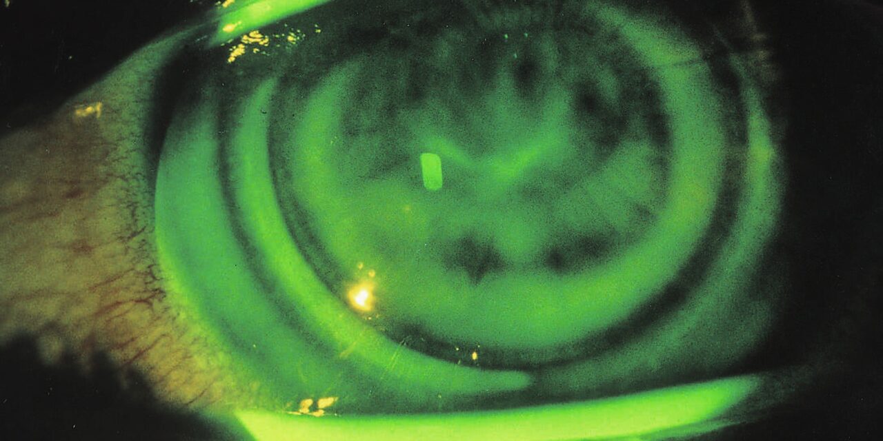
Contact lens-induced corneal warpage is a reversible alteration of normal corneal shape caused by chronic contact lens wear, most classically associated with older, poorly fitted, or long-worn rigid lenses, and it is a genuinely important condition to recognize because it can convincingly mimic keratoconus, discussed in its own dedicated article on this site, on topography.
The critical distinguishing feature is reversibility: unlike keratoconus, a progressive, permanent structural disease, warpage resolves gradually once the causative lens wear stops, which is exactly the property that makes recognizing it correctly, rather than mistaking it for keratoconus, so clinically important.
Understanding this distinction matters most acutely before refractive surgery, discussed in relation to LASIK in its own dedicated article on this site, where mistaking warpage for early keratoconus, or vice versa, has significant and very different implications for surgical candidacy.
Mechanism
Chronic mechanical pressure from a rigid contact lens, particularly one with a poor fit relative to the individual cornea’s own curvature, can gradually mold and reshape the anterior corneal surface over months to years of regular wear.
This molding effect is a physical, mechanical phenomenon rather than a true structural or biochemical disease process, which is precisely why the cornea gradually returns toward its natural shape once the causative mechanical pressure from lens wear is removed.
Soft lenses can occasionally cause a milder version of this same phenomenon, though clinically significant warpage is classically and most commonly associated with rigid gas-permeable or older hard contact lens wear.
Clinical and Topographic Findings
- Irregular astigmatism on topography, sometimes with an asymmetric or skewed pattern that can closely resemble the topographic signature of early keratoconus
- Reduced best-corrected visual acuity with spectacles, reflecting the irregular corneal surface, though contact lenses themselves often still provide reasonably good vision by effectively masking the irregularity
- A pattern of topographic change, when serial measurements are available, that correlates with the specific lens wear pattern and improves progressively once lens wear is discontinued
- Absence of other classic keratoconus signs, including Vogt’s striae, a Fleischer ring, or significant progressive corneal thinning, on careful slit-lamp exam
All-fit Slit-Lamp Adapter
Record and share exactly what you see at the slit lamp. One adapter fits any slit lamp or surgical microscope — and any smartphone.
From Choroida — the team behind this siteDistinguishing Warpage From Keratoconus
The single most useful diagnostic step is discontinuing contact lens wear and repeating corneal topography after an adequate washout period, since true warpage shows progressive improvement and eventual normalization, while keratoconus does not improve and may continue its own independent, unrelated progression.
The required washout period varies depending on lens type and duration of wear, ranging from days for recent, mild soft lens wear to many weeks or even months for longstanding rigid lens wear, and topography needs to be repeated at intervals during this washout to confirm a genuine trend toward stabilization or normalization.
Corneal thickness measurements and posterior corneal surface topography, less affected by anterior lens-induced molding than anterior curvature alone, can provide additional supportive information when the anterior topographic picture remains ambiguous.
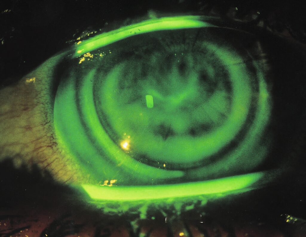
Why This Matters Before Refractive Surgery
Proceeding with LASIK or another refractive procedure based on topography still affected by warpage risks two serious errors: operating on an eye with a temporarily distorted, inaccurate topographic map, or missing a case of genuine early keratoconus masked by lens-induced changes that happen to be improving in the same direction.
Standard refractive surgery screening protocols specifically require an adequate contact lens washout period with topographic stability confirmed on serial measurements before finalizing surgical candidacy, precisely because of this diagnostic overlap.
Patients need clear counseling about why this washout period, sometimes lasting weeks to months depending on their lens type and wear history, is a necessary part of accurate preoperative evaluation rather than an arbitrary delay.
Management
Discontinuing the causative lens wear and switching to spectacles or a different lens modality during the recovery period allows the cornea to gradually return toward its natural shape.
Serial topography during this recovery period documents the expected trend toward normalization, confirming the diagnosis retrospectively as the cornea stabilizes.
Once topographic stability is confirmed, whether for the purpose of clearing a patient for refractive surgery or simply resolving the underlying diagnostic uncertainty, normal contact lens wear, ideally with a better-fitted lens design, can generally be resumed if desired.


Document what you see
Two smartphone imaging tools built for everyday clinic use — one for the slit lamp, one for the fundus.
From Choroida — the team behind this siteReferences
- Wilson SE, Lin DT, Klyce SD, et al. Rigid contact lens decentration: a risk factor for corneal warpage. CLAO Journal.
- American Academy of Ophthalmology. Basic and Clinical Science Course, Section 13: Refractive Surgery.
- Ruiz-Montenegro J, Mafra CH, Wilson SE, et al. Corneal topographic changes after discontinuation of rigid contact lens wear. Ophthalmology.
Test yourself
A few questions straight from this article.
-
Which feature separates contact lens-induced corneal warpage from keratoconus?
Warpage resolves gradually after the causative lens wear is discontinued, whereas keratoconus is a progressive, permanent structural disease. Reversibility is the critical distinguishing feature. -
What mechanism produces contact lens-induced corneal warpage?
Sustained mechanical pressure from a lens, especially one fitted poorly to the individual cornea, gradually moulds the anterior surface over months to years. The effect is physical, not a true disease process. -
Which lens type is classically associated with clinically significant corneal warpage?
Soft lenses can occasionally cause a milder version, but clinically significant warpage is classically associated with rigid gas-permeable or older hard lens wear. -
What visual acuity pattern is typical in contact lens-induced corneal warpage?
The irregular surface reduces best-corrected spectacle acuity, but contact lenses themselves often still give reasonably good vision by masking that irregularity. -
Which slit-lamp signs are characteristically absent in contact lens-induced corneal warpage?
Careful slit-lamp examination in warpage shows an absence of the classic keratoconus signs, including Vogt's striae, a Fleischer ring, and significant progressive corneal thinning. -
What is the single most useful step for distinguishing corneal warpage from keratoconus?
True warpage shows progressive improvement and eventual normalisation once lens wear stops, while keratoconus does not improve and may continue its own independent progression. -
How long may the washout period need to be after longstanding rigid contact lens wear?
The washout ranges from days after recent, mild soft lens wear to many weeks or even months after longstanding rigid lens wear, with topography repeated at intervals throughout. -
Which measurements are less affected by anterior lens-induced moulding and can help when topography is ambiguous?
Corneal thickness measurements and posterior corneal surface topography are less affected by anterior moulding than anterior curvature alone, so they give supportive information when the picture is unclear. -
What risk arises from planning refractive surgery on topography still affected by warpage?
Operating on warped topography risks either treating a temporarily distorted, inaccurate map or missing genuine early keratoconus hidden by lens-induced changes moving in the same direction. -
What can a patient with resolved corneal warpage do once topographic stability is confirmed?
Once serial topography confirms stability, normal contact lens wear can generally be resumed if the patient wishes, preferably with a better-fitted lens design.
