
Overview
Progressive Retinal Atrophy (PRA) is a group of genetic diseases characterized by the progressive degeneration of the retina, leading to vision loss and eventual blindness in animals.

PRA affects various species, including dogs, cats, and horses, and is one of the most common inherited retinal disorders in veterinary medicine.
This article provides a comprehensive overview of PRA in animals, covering its disease entity, causes, clinical signs, diagnosis, treatment options, and prognosis.
Progressive Retinal Atrophy (PRA) Disease Entity
Progressive Retinal Atrophy (PRA) encompasses a heterogeneous group of inherited retinal degenerative disorders characterized by the gradual deterioration of retinal function and structure.
PRA primarily affects the photoreceptor cells of the retina, namely the rods and cones, leading to progressive vision loss over time.
While PRA can manifest in various forms and progressions depending on the species and genetic mutation involved, the ultimate outcome is bilateral blindness in affected animals.
Causes
PRA is primarily caused by genetic mutations inherited in an autosomal recessive, autosomal dominant, or X-linked manner, depending on the specific genetic mutation involved.
These mutations disrupt the normal function and structure of retinal cells, leading to their progressive degeneration.
While the exact genetic mutations responsible for PRA vary among different animal species and breeds, genetic testing has identified several gene mutations associated with the disease, facilitating early detection and breeding strategies to reduce the prevalence of PRA in susceptible populations.
Progressive Retinal Atrophy (PRA) Clinical Signs
Clinical signs of Progressive Retinal Atrophy (PRA) typically manifest gradually and progress over time as the disease advances.
Common clinical signs observed in affected animals include:
- Night blindness (nyctalopia): Affected animals may exhibit difficulty seeing in low-light conditions, particularly during dusk or nighttime, due to the initial degeneration of rod photoreceptor cells responsible for dim light vision.
- Loss of visual acuity: As PRA progresses, affected animals may experience a decline in visual acuity, leading to impaired vision during daylight hours and difficulty navigating their surroundings.
- Pupillary abnormalities: PRA may also be associated with abnormal pupillary light reflexes, including sluggish or absent responses to light stimulation, indicating dysfunction of retinal cells involved in regulating pupil size.
- Changes in behavior: Affected animals may display behavioral changes indicative of visual impairment, such as reluctance to navigate unfamiliar environments, bumping into objects, or becoming disoriented.
Diagnosis
Diagnosing Progressive Retinal Atrophy (PRA) in animals typically involves a combination of clinical evaluation, ophthalmic examination, and genetic testing.
Diagnostic steps may include:
- Ophthalmic examination: A comprehensive ophthalmic evaluation by a qualified veterinarian or veterinary ophthalmologist, including assessment of visual function, funduscopic examination to evaluate retinal health and integrity, and electroretinography (ERG) to assess retinal function and detect abnormalities characteristic of PRA.
- Genetic testing: Molecular genetic testing to identify specific gene mutations associated with PRA in affected animals, facilitating early detection, breeding decisions, and genetic counseling for susceptible breeds.
Progressive Retinal Atrophy (PRA) Treatment
Currently, there is no definitive treatment to reverse or halt the progression of Progressive Retinal Atrophy (PRA) in animals.
Management primarily focuses on supportive care and strategies to optimize the quality of life for affected animals. Symptomatic treatment options may include:
- Environmental modifications: Creating a safe and familiar environment for visually impaired animals by minimizing hazards, maintaining consistent routines, and providing tactile or auditory cues to aid navigation.
- Nutritional supplements: Some veterinarians may recommend nutritional supplements containing antioxidants, omega-3 fatty acids, or other beneficial nutrients to support retinal health and potentially slow the progression of PRA, although their efficacy remains uncertain.
- Behavioral adaptation: Implementing behavioral training techniques to facilitate orientation and mobility in visually impaired animals, including scent-based navigation, tactile cues, and auditory commands.
Prognosis
The prognosis for animals diagnosed with Progressive Retinal Atrophy (PRA) varies depending on the specific genetic mutation involved, disease progression, and individual factors.
While PRA ultimately leads to bilateral blindness in affected animals, the rate of disease progression and impact on the animal’s quality of life can vary.
Early detection, proactive management, and supportive care are essential in optimizing the quality of life for animals affected by this degenerative retinal disorder.
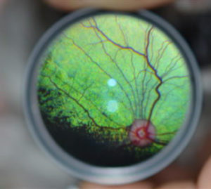
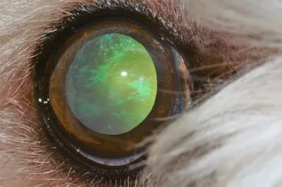
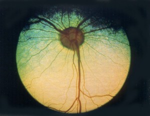
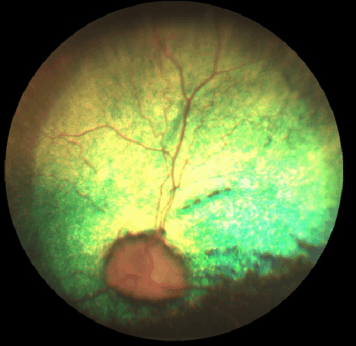
Would you have interest in taking retinal images with your smartphone?
Fundus photography is superior to fundus analysis as it enables intraocular pathologies to be photo-captured and encrypted information to be shared with colleagues and patients.
Recent technologies allow smartphone-based attachments and integrated lens adaptors to transform the smartphone into a portable fundus camera and Retinal imaging by smartphone.
RETINAL IMAGING BY YOUR SMARTPHONE
References
- Acland, G. M., Aguirre, G. D., Ray, J., Zhang, Q., Aleman, T. S., Cideciyan, A. V., … & Hauswirth, W. W. (2001). Gene therapy restores vision in a canine model of childhood blindness. Nature Genetics, 28(1), 92-95.
- Aguirre, G. D., Rubin, L. F., & Schmehl, D. M. (1978). Clinical manifestations of inherited retinal degeneration in the dog. Investigative Ophthalmology & Visual Science, 17(7), 838-847.
- Beltran, W. A., & Aguirre, G. D. (2009). Canine Retinal Degenerations: Clinical, Histological, and Genetic Investigations. In Small Animal Ophthalmic Atlas and Guide (pp. 232-244). John Wiley & Sons.
- Miyadera, K., Acland, G. M., & Aguirre, G. D. (2012). Genetic and phenotypic variations of inherited retinal diseases in dogs: the power of within-and across-breed studies. Mammalian Genome, 23(1-2), 40-61.
RETINAL IMAGING BY YOUR SMARTPHONE

RETINAL IMAGING BY YOUR SMARTPHONE