
An intumescent cataract is a lens that has absorbed enough fluid to swell significantly, and this single structural fact, a lens that is now larger and under more internal pressure than a typical cataract, is what drives both of its major clinical consequences: a real risk of angle closure before surgery and a specific, well-recognized set of technical hazards once surgery begins.
What Makes a Cataract “Intumescent”
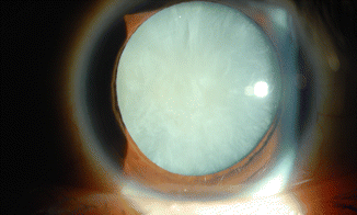
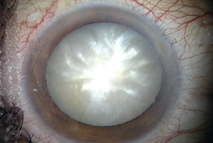
As some cataracts mature, the lens can take up water and swell, increasing significantly in anteroposterior thickness and overall volume, a process that can happen relatively quickly compared with the typically slow, gradual progression of an ordinary age-related cataract.
This swelling is not simply a cosmetic or incidental change, since a larger, more swollen lens pushes the iris-lens diaphragm forward, crowding the anterior chamber, and it also creates elevated pressure within the lens capsule itself, both of which have direct, practical consequences for the patient and the surgeon.
The Angle Closure Risk
When an intumescent lens pushes the iris forward enough to crowd the anterior chamber angle, it can precipitate phacomorphic glaucoma, a specific form of acute angle closure caused directly by the lens itself rather than by the anatomic predisposition that underlies primary angle closure (see phacomorphic glaucoma for a detailed look at this presentation and its emergency management).
Recognizing an intumescent cataract, particularly in a patient presenting with pain, redness, and blurred vision, should prompt an urgent check of intraocular pressure and anterior chamber depth, since this combination can evolve into a true ophthalmic emergency.
All-fit Slit-Lamp Adapter
Record and share exactly what you see at the slit lamp. One adapter fits any slit lamp or surgical microscope — and any smartphone.
From Choroida — the team behind this siteClinical Recognition
- A visibly swollen, often densely white lens on examination, sometimes with the anterior capsule appearing tense or under pressure
- A shallower anterior chamber than would be expected for the patient’s age or refractive history, particularly when compared with the fellow eye
- Rapid, sometimes recent-onset, dense visual loss reported by the patient, reflecting how quickly some intumescent cataracts can develop compared with typical age-related progression
- Elevated intraocular pressure, when phacomorphic glaucoma has already developed
Why Intumescent Cataracts Complicate Surgery
The elevated internal pressure within a swollen lens creates a specific, well-recognized intraoperative hazard: at the moment the anterior capsule is opened to begin a capsulorhexis, this internal pressure can cause the capsulotomy to run outward uncontrollably in a sudden radial tear, a complication significant enough that intumescent cataracts warrant a modified surgical approach from the outset rather than a standard technique.
- A dense, white, mature cortex can also make it difficult to visualize and safely complete the capsulorhexis using standard technique alone, since the normal red reflex that guides capsulorhexis creation is often absent or severely degraded
Modified Surgical Technique
Trypan Blue Capsule Staining
Staining the anterior capsule with trypan blue dye improves visualization of the capsulorhexis margin in the absence of a useful red reflex, a technique particularly valuable in white, mature, and intumescent cataracts where standard illumination alone is often inadequate.
Decompressing the Capsular Bag Before Capsulorhexis
A specific technique used for intumescent cataracts involves first aspirating a small amount of liquefied cortex through a small initial puncture in the anterior capsule, deliberately decompressing the pressurized capsular bag before completing the full capsulorhexis, which substantially reduces the risk of an uncontrolled radial tear.
Careful, Controlled Capsulorhexis Technique
Even with decompression, a smaller initial capsulorhexis, careful use of high-viscosity ophthalmic viscosurgical devices to maintain a stable chamber, and a slower, more controlled technique throughout are generally used for these cases, reflecting the overall higher-risk nature of the capsulorhexis step in an intumescent lens.
Managing Coexisting Phacomorphic Glaucoma
When an intumescent cataract has already caused acute angle closure, initial pressure-lowering measures are used to stabilize the eye and clear corneal edema before proceeding to definitive cataract surgery, following the same emergency management principles used for phacomorphic glaucoma generally, since cataract extraction itself is the definitive treatment once the eye is adequately prepared for safe surgery.
Preoperative Counseling
Given the recognized additional technical risk, patients with an intumescent cataract benefit from specific counseling about the modestly higher complication risk of their surgery compared with a routine, non-swollen cataract, along with an explanation of why a modified surgical approach, including capsule staining and a decompression step, will be used.
Prognosis
With recognition of the intumescent nature of the cataract before surgery and use of the specific modified techniques described above, the great majority of these cases are managed safely with outcomes comparable to routine cataract surgery.
The main driver of complications in this setting is failure to anticipate the intumescent lens and its specific risks, proceeding with standard technique as though the case were routine, which is why identifying this specific cataract morphology preoperatively is a genuinely consequential part of surgical planning (see cataract morphology classification for the broader framework this specific pattern fits within).
Related Reading


Document what you see
Two smartphone imaging tools built for everyday clinic use — one for the slit lamp, one for the fundus.
From Choroida — the team behind this siteReferences
- Vasavada AR, Praveen MR, Tassignon MJ, et al. Subclinical capsulorhexis anomalies in phacoemulsification of white intumescent cataracts. J Cataract Refract Surg. 2009;35:298-302.
- Chakrabarti A, Singh S, Krishnadas R. Phacoemulsification in eyes with white cataract. J Cataract Refract Surg. 2000;26:1041-1047.
- Marques FF, Marques DM, Osher RH, Osher JM. Fate of anterior capsule tears during cataract surgery. J Cataract Refract Surg. 2006;32:1638-1642.
- Prajna NV, Lalitha P, Mahalakshmi R, Prajna L. A randomised trial comparing pre-operative povidone iodine 5% and trypan blue as an adjunct to cataract surgery. Br J Ophthalmol. 2010;94:1613-1615.
