
Granular corneal dystrophy is a bilateral, autosomal dominant stromal dystrophy characterized by discrete, sharply demarcated, breadcrumb- or snowflake-like deposits of hyaline material within the anterior-to-mid corneal stroma, separated by clear intervening stroma.
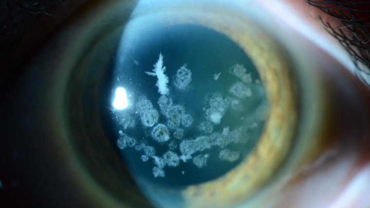
This is a pattern distinct enough from the other classic corneal stromal dystrophies that the three (granular, lattice, and macular) are traditionally taught together specifically to reinforce how differently each one looks despite superficially similar underlying territory.
Genetics and Classification
Granular corneal dystrophy, like lattice type I, results from mutations in TGFBI.
However, the specific mutation determines the resulting clinical phenotype: classic granular dystrophy (sometimes designated type I) shows the discrete, well-demarcated hyaline deposits described above.
Meanwhile, a related but distinct phenotype — Avellino corneal dystrophy (granular-lattice dystrophy, type II) — results from a different TGFBI mutation and produces a combined picture with features of both granular deposits and lattice-type linear lesions in the same cornea.
This shared genetic origin across several distinct-appearing corneal dystrophies is a useful illustration of how a single gene can produce different phenotypes depending on the exact mutation involved, which is why genetic testing, when pursued, needs to characterize the specific mutation rather than simply confirming involvement of the TGFBI gene in general.
Clinical Presentation
The condition typically becomes apparent in the first or second decade of life, though visual symptoms are often mild for a long time, because the deposits characteristically spare the periphery and the intervening stroma between deposits remains genuinely clear.
This is a meaningful practical difference from lattice or macular dystrophy, where progressive diffuse stromal haze more consistently and more significantly impairs vision.
Many patients with granular dystrophy retain good vision for decades, with visual impact becoming more significant only later in life as deposits gradually become more numerous, larger, and occasionally begin to coalesce, a genuinely reassuring long-term trajectory relative to some of the other stromal dystrophies.
All-fit Slit-Lamp Adapter
Record and share exactly what you see at the slit lamp. One adapter fits any slit lamp or surgical microscope — and any smartphone.
From Choroida — the team behind this siteExam Findings
- Discrete, sharply demarcated, white-gray, breadcrumb- or snowflake-shaped deposits within the anterior-to-mid stroma, most concentrated centrally
- Clear cornea between individual deposits, at least in earlier stages of disease — a key distinguishing feature from the diffuse interlesional haze of lattice dystrophy and the confluent haze of macular dystrophy
- Deposits typically increase in number and can enlarge or begin to coalesce with age, sometimes eventually reducing the clarity of the previously unaffected intervening stroma in more advanced, longstanding disease
- Preserved corneal sensation in most cases, unlike some other corneal conditions where reduced sensation is a prominent feature
- Recurrent corneal erosion can occur, though generally less prominently and less frequently than in lattice dystrophy
Differential Diagnosis
- Lattice corneal dystrophy — branching, linear, refractile deposits with diffuse interlesional haze, rather than the discrete, well-demarcated deposits with clear intervening stroma typical of granular dystrophy, discussed in its own dedicated article on this site
- Macular corneal dystrophy — diffuse, cloudy stromal opacification without discrete deposits, and notably autosomal recessive rather than dominant, distinguishing its inheritance pattern from both granular and lattice dystrophy
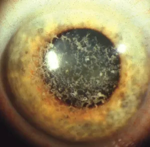
- Avellino (granular-lattice) corneal dystrophy — a combined phenotype from a related TGFBI mutation, showing features of both classic patterns simultaneously in the same cornea
- Climatic droplet keratopathy — environmentally acquired rather than inherited, with a characteristic interpalpebral distribution and relevant occupational/environmental exposure history, discussed in its own dedicated article on this site
A Practical Surgical Note: Avellino Dystrophy and LASIK
A specifically important clinical pearl involves Avellino corneal dystrophy: patients with this condition, if the diagnosis is unrecognized before refractive surgery, have experienced dramatic, rapid worsening of stromal deposits following LASIK, thought to be triggered by the corneal wound-healing response to the procedure.
This is a genuinely practical reason to specifically screen for any family history of corneal dystrophy, and to examine the cornea carefully for subtle deposits, before performing refractive surgery in any patient.
A missed, subclinical case can have significantly worse consequences after LASIK than it would have had if simply left untreated, which is exactly why a careful preoperative slit-lamp exam and family history remain essential even for a patient who otherwise seems like an ideal refractive surgery candidate.
Management
Many patients with granular dystrophy require no treatment for extended periods, given the preserved central clarity in earlier disease, with management limited to lubrication and monitoring.
As deposits progress and begin to significantly affect vision, phototherapeutic keratectomy can remove more superficial anterior stromal deposits and improve vision, though, as with lattice dystrophy, deposits characteristically recur over subsequent years since the underlying genetic tendency toward abnormal protein deposition is not altered by removing existing material.
Corneal transplantation (penetrating or deep anterior lamellar keratoplasty) is reserved for more advanced disease with significant visual impact, and, similar to lattice dystrophy, recurrence of granular deposits within the graft over subsequent years is a recognized long-term consideration that patients should be counseled about at the time surgery is planned.
Because both surgical options — phototherapeutic keratectomy and transplantation — offer only a temporary reprieve rather than a cure, patients benefit from understanding early on that management of this condition is a long-term, ongoing process rather than a single definitive intervention.


Document what you see
Two smartphone imaging tools built for everyday clinic use — one for the slit lamp, one for the fundus.
From Choroida — the team behind this siteReferences
- Klintworth GK. Corneal dystrophies. Orphanet Journal of Rare Diseases.
- Afshari NA, Mullally JE, Afshari MA, et al. Survey of patients with granular, lattice, avellino, and Reis-Bücklers corneal dystrophies for mutations in the BIGH3 and gelsolin genes. Archives of Ophthalmology.
- Jun RM, Tchah H, Kim TI, et al. Avellino corneal dystrophy after LASIK. Ophthalmology.
- American Academy of Ophthalmology. Basic and Clinical Science Course, Section 8: External Disease and Cornea.
Test yourself
A few questions straight from this article.
-
Which material forms the deposits of granular corneal dystrophy?
Granular dystrophy is characterised by discrete, sharply demarcated breadcrumb- or snowflake-like deposits of hyaline material within the anterior-to-mid corneal stroma. -
Mutations in which gene cause granular corneal dystrophy?
Granular corneal dystrophy results from TGFBI mutations, the same gene responsible for lattice type I, with the specific mutation determining which phenotype appears. -
What corneal appearance characterises Avellino corneal dystrophy?
Avellino (granular-lattice) dystrophy arises from a different TGFBI mutation and produces a combined picture showing both granular deposits and lattice-type linear lesions in the same cornea. -
Why must genetic testing in TGFBI corneal dystrophy identify the specific mutation?
A single gene can produce several distinct-appearing dystrophies depending on the exact mutation, so testing must characterise the specific mutation rather than just confirm TGFBI involvement. -
Why do patients with granular corneal dystrophy often keep good vision for decades?
Visual symptoms stay mild for a long time because the deposits characteristically spare the periphery and the stroma between them remains genuinely clear. -
What is the typical state of corneal sensation in granular corneal dystrophy?
Corneal sensation is preserved in most cases of granular dystrophy, unlike some other corneal conditions in which reduced sensation is a prominent feature. -
Which inheritance pattern does macular corneal dystrophy follow, unlike granular dystrophy?
Macular corneal dystrophy is autosomal recessive, which separates its inheritance pattern from the autosomal dominant granular and lattice dystrophies. -
Which feature identifies climatic droplet keratopathy rather than granular corneal dystrophy?
Climatic droplet keratopathy is environmentally acquired rather than inherited, with a characteristic interpalpebral distribution and a relevant occupational or environmental exposure history. -
What happens to unrecognised Avellino corneal dystrophy after LASIK?
Patients with unrecognised Avellino dystrophy have experienced dramatic, rapid worsening of stromal deposits after LASIK, thought to be triggered by the corneal wound-healing response, which is why family history and careful slit-lamp exam matter before refractive surgery. -
What should patients expect after phototherapeutic keratectomy for granular corneal dystrophy?
Phototherapeutic keratectomy removes superficial anterior stromal deposits and improves vision, but deposits characteristically recur because the underlying genetic tendency to deposit protein is unchanged.
