
Plaque brachytherapy delivers radiation to a choroidal melanoma from a small radioactive disc sutured to the sclera, and it is the most widely used eye-preserving treatment for medium and small uveal melanomas.
The Collaborative Ocular Melanoma Study showed that it gave survival equivalent to enucleation for medium tumors, which changed practice worldwide.
The tumor is usually controlled, but the eye pays a price in radiation damage to the retina and optic nerve, and the patient still needs lifelong surveillance for metastasis.
Choroidal Melanoma and the Role of Plaque Brachytherapy
Choroidal melanoma is the most common primary intraocular malignancy in adults.
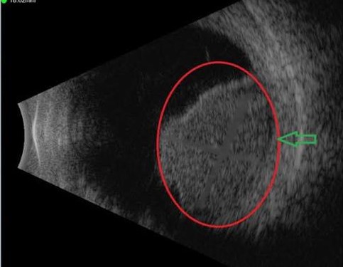
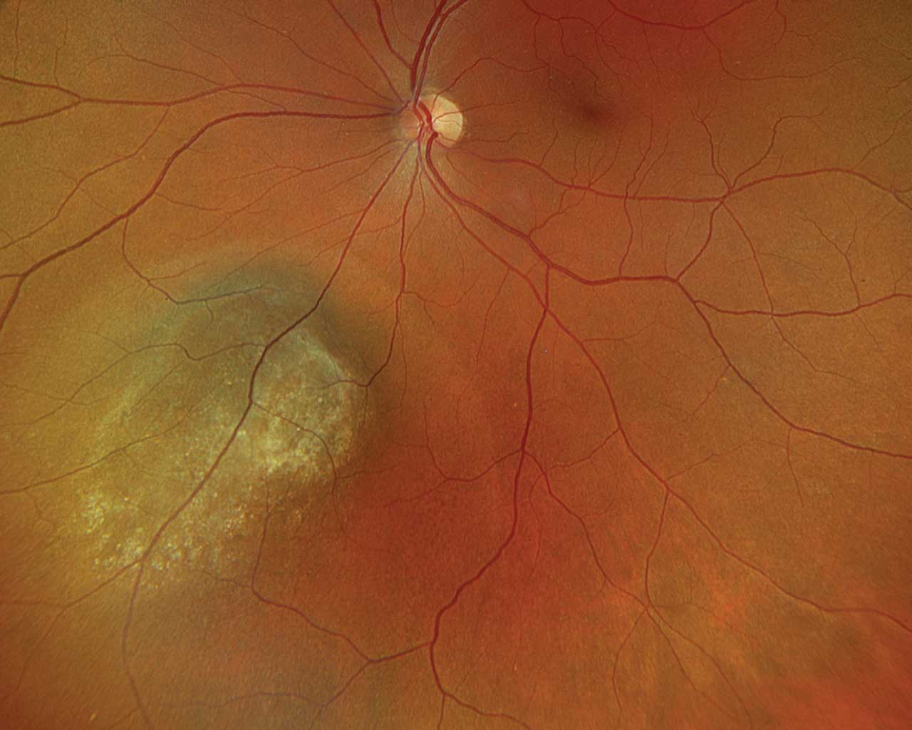
It is diagnosed by clinical examination and imaging, with ultrasonography showing a dome- or mushroom-shaped mass with low internal reflectivity, and OCT and autofluorescence documenting subretinal fluid and orange pigment (see choroidal nevus versus small melanoma).
Treatment aims are local tumor control, preservation of the eye and vision when possible, and reduction of the risk of metastasis, which occurs mainly in the liver.
Options include plaque brachytherapy, proton beam radiotherapy, enucleation, local resection, and, for small lesions, observation or laser-based treatments.
Evidence From the COMS
The COMS randomized patients with medium choroidal melanoma (apical height 2.5 to 10 mm and basal diameter up to 16 mm) to iodine-125 plaque therapy or enucleation.
Survival was similar between the two groups over 12 years of follow-up, and enucleation did not improve survival compared with brachytherapy.
Local treatment failure requiring enucleation occurred in roughly one in ten eyes within five years.
Visual acuity outcomes showed that about two in five eyes lost substantial acuity within three years, which reflects radiation effects on the macula and optic nerve.
The findings established plaque brachytherapy as the standard of care for medium tumors, and for suitable small tumors.
Fundus Explorer Pro
Photograph the retinal findings described here with the phone already in your pocket — 22 D optics and built-in illumination in one handheld unit.
From Choroida — the team behind this siteTechnique
Isotopes
- Iodine-125 is the most common in North America and was used in the COMS
- Ruthenium-106 is a beta emitter with a shallow penetration, suited to tumors of limited thickness, and widely used in Europe
- Palladium-103 is used in some centers
Planning and Delivery
Dose calculation uses tumor dimensions, plaque size, and the position of the tumor relative to the macula and optic disc.
In the COMS, the prescribed dose was 85 Gy to the tumor apex.
The plaque is sewn to the sclera over the tumor after localization, and it remains in place for several days, then is removed in a second procedure.
Notched or custom plaques treat tumors near the optic disc.
Adjuncts
Transpupillary thermotherapy and laser have been used as adjuncts in some centers.
Anti-VEGF injections and intravitreal steroids have been used to reduce the rate of radiation maculopathy, although evidence is limited.
Radiation Complications
Radiation effects appear months to years after treatment and depend on dose and location.
- Radiation retinopathy with microangiopathy, macular edema, and ischemia is the leading cause of visual loss (see radiation retinopathy)
- Radiation optic neuropathy produces disc swelling followed by atrophy (see radiation optic neuropathy)
- Cataract, especially with anterior tumors
- Neovascular glaucoma from retinal ischemia and tumor necrosis (see neovascular glaucoma)
- Vitreous hemorrhage
- Scleral thinning or necrosis under the plaque
- Diplopia from muscle damage
- Dry eye and eyelid effects
Regular follow-up detects these complications while they are treatable, and anti-VEGF injections, steroids, laser, and surgery are used as needed.
Surveillance and Metastasis
The main threat to life is metastatic disease, which develops in a substantial proportion of patients, most often in the liver, and sometimes years after successful local treatment.
Prognostic tumor testing with gene expression profiling or chromosome 3 and BAP1 analysis can identify high-risk tumors.
Surveillance usually combines periodic liver imaging with ultrasound or MRI and liver function testing, at intervals set by the risk category.
For metastatic uveal melanoma, tebentafusp improved overall survival compared with investigator’s choice therapy in HLA-A*02:01-positive patients.
Patients need to understand that local control does not remove the risk of metastasis, and that follow-up is lifelong.
Counseling Before Treatment
Patients should hear four messages before plaque therapy.
- The aim is to control the tumor and keep the eye, and the decision does not change the risk of metastasis, which depends mainly on tumor biology
- Vision will probably decline over the next few years if the tumor is near the macula or optic disc, and early treatment of radiation complications may help
- The plaque is temporary, and radiation safety measures for family contact are explained by the treating team
- Follow-up is lifelong, with both eye examinations and systemic surveillance
Psychological support and contact with a specialist ocular oncology service are valuable at the time of diagnosis.
Local Recurrence
Local recurrence appears as tumor growth on ultrasound or a new area of elevated pigment or fluid.
It is managed with repeat plaque, proton beam therapy, resection, or enucleation.
The risk is higher with large tumors, tumors near the disc, and inadequate dose.
Prognosis and Visual Outcome
Local tumor control is achieved in most patients, and many keep the eye.
Vision often declines over several years from radiation maculopathy and optic neuropathy, particularly for tumors near the macula or optic disc.
Better preservation is seen with tumors distant from the posterior pole.
Patients should be offered rehabilitation and support, and early treatment of macular edema may preserve vision.


Document what you see
Two smartphone imaging tools built for everyday clinic use — one for the slit lamp, one for the fundus.
From Choroida — the team behind this siteReferences
- Collaborative Ocular Melanoma Study Group. The COMS randomized trial of iodine 125 brachytherapy for choroidal melanoma: V. Twelve-year mortality rates and prognostic factors: COMS report No. 28. Arch Ophthalmol. 2006;124:1684-1693.
- Melia BM, Abramson DH, Albert DM, et al. Collaborative ocular melanoma study randomized trial of I-125 brachytherapy for medium choroidal melanoma. I. Visual acuity after 3 years: COMS report no. 16. Ophthalmology. 2001;108:348-366.
- Jampol LM, Moy CS, Murray TG, et al. The COMS randomized trial of iodine 125 brachytherapy for choroidal melanoma: IV. Local treatment failure and enucleation in the first 5 years after brachytherapy. COMS report no. 19. Ophthalmology. 2002;109:2197-2206.
- Shields CL, Cater J, Shields JA, et al. Plaque radiotherapy for uveal melanoma: long-term visual outcome in 1106 consecutive patients. Arch Ophthalmol. 2000;118:1219-1228.
- Nathan P, Hassel JC, Rutkowski P, et al. Overall survival benefit with tebentafusp in metastatic uveal melanoma. N Engl J Med. 2021;385:1196-1206.
- Jager MJ, Shields CL, Cebulla CM, et al. Uveal melanoma. Nat Rev Dis Primers. 2020;6:24.
