
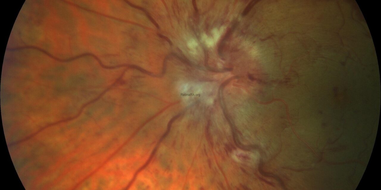
Papillophlebitis — also called optic disc vasculitis or, in older literature, “big blind spot syndrome” — is a condition affecting young, otherwise healthy adults that sits in an uncomfortable diagnostic space between optic disc edema and central retinal vein occlusion, sharing features of both without cleanly fitting either category.
Recognizing it as its own entity matters because its prognosis and workup differ meaningfully from a typical CRVO in an older patient with vascular risk factors.
What Distinguishes Papillophlebitis
Unlike classic central retinal vein occlusion, which predominantly affects older patients with hypertension, diabetes, or glaucoma, papillophlebitis typically presents in patients under 50, often without any significant systemic vascular risk factors.
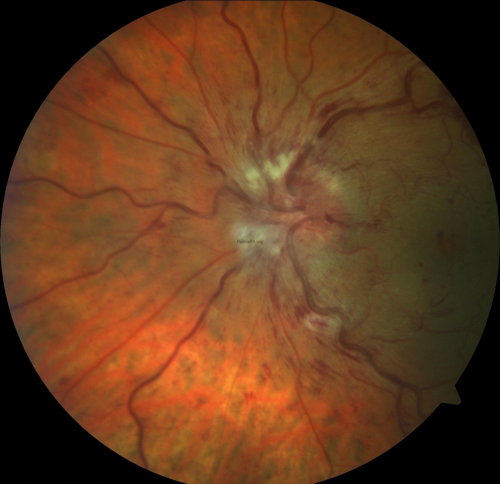
The fundus appearance overlaps substantially with mild to moderate non-ischemic CRVO — disc edema, venous dilation and tortuosity, scattered retinal hemorrhages — but the degree of vascular compromise is generally milder, and the visual prognosis is correspondingly better than typical CRVO in most cases.
The name reflects an older hypothesis that inflammation of the central retinal vein itself (a true “phlebitis”) underlies the condition, though this has never been definitively confirmed histopathologically, and the exact pathophysiology remains incompletely understood.
Some authors view it as a mild, self-limited variant of non-ischemic CRVO in a younger population rather than a distinct vasculitic process.
This ongoing uncertainty about whether papillophlebitis is truly a separate disease or simply the young end of the CRVO spectrum is reflected in the lack of a single, universally agreed diagnostic test or treatment protocol, making careful clinical judgment particularly important in each individual case.
Clinical Presentation
Patients typically present with mild to moderate, often unilateral, painless vision loss or blurring, sometimes accompanied by a visual field defect corresponding to the enlarged blind spot that gave the condition its older descriptive name.
Vision loss is generally less severe than in ischemic CRVO, and many patients retain relatively good visual acuity even at presentation.
Because affected patients are often young, healthy, and free of the usual vascular risk factors that might otherwise raise concern, a new painless visual disturbance in this population can be initially dismissed or attributed to a benign cause, making a careful dilated fundus exam an important step even when the presentation seems unlikely to reflect serious retinal vascular disease.
Fundus Explorer Pro
Photograph the retinal findings described here with the phone already in your pocket — 22 D optics and built-in illumination in one handheld unit.
From Choroida — the team behind this siteExam Findings
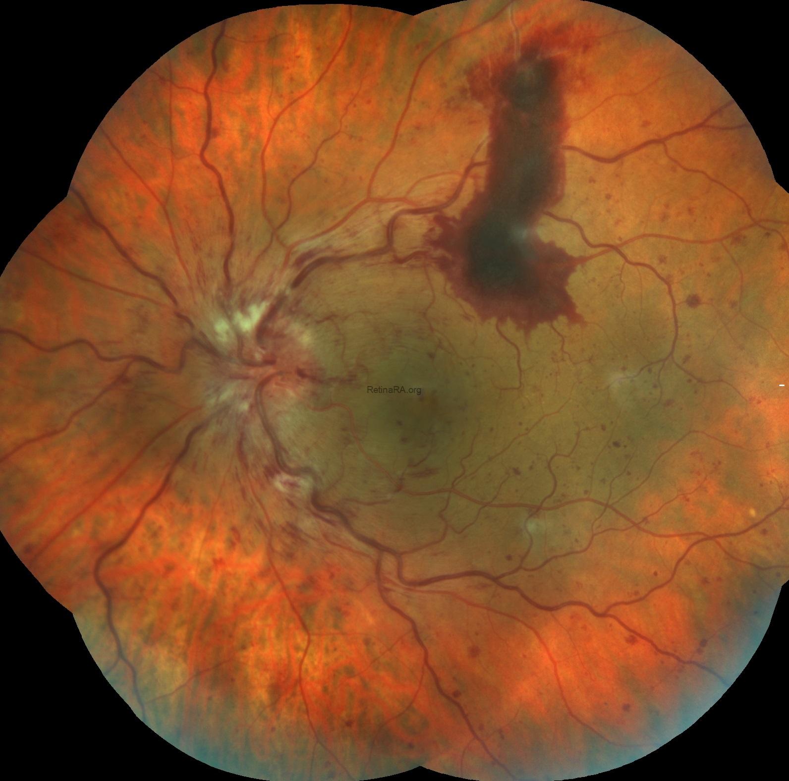
- Optic disc edema, often prominent
- Dilated, tortuous retinal veins
- Scattered retinal hemorrhages, typically less extensive than in ischemic CRVO
- Absence of significant retinal ischemia on fluorescein angiography (a key distinguishing feature from ischemic CRVO)
- Relatively preserved visual acuity relative to the degree of fundus findings
Differential Diagnosis
- Non-ischemic central retinal vein occlusion — the closest mimic, distinguished mainly by patient age and the absence of typical vascular risk factors in papillophlebitis
- Papilledema from raised intracranial pressure — typically bilateral, associated with headache and other signs of elevated ICP, without the same degree of venous dilation and hemorrhage
- Anterior ischemic optic neuropathy — more acute, more significant vision loss, altitudinal field defect, different demographic (often older, or younger with specific risk factors)
- Optic neuritis — pain with eye movement, more prominent color vision loss, distinct MRI findings
Fluorescein angiography is particularly useful for this distinction, since papillophlebitis characteristically lacks the extensive capillary non-perfusion seen in ischemic CRVO — the single most important prognostic branch point in the vein occlusion spectrum generally.
Workup
Despite the relative rarity of identifiable systemic vascular risk factors, a reasonable baseline evaluation includes blood pressure measurement, basic metabolic and hematologic screening, and consideration of a hypercoagulable workup in younger patients, particularly if there is a personal or family history of thrombosis.
Because the entity by definition occurs without the risk factors typical of older CRVO patients, some clinicians pursue a broader initial workup rather than they would for a typical vein occlusion in an older patient, though there is no universally agreed-upon protocol, and the intensity of workup is often tailored to the individual patient’s history and any atypical features present.
Management
Most cases are managed conservatively with observation, given the generally favorable natural history and relatively good baseline vision.
Systemic corticosteroids have been used in some cases based on the presumed inflammatory component, though evidence for this approach is limited to small case series rather than controlled trials, and practice varies between clinicians, with some reserving steroids for cases with more pronounced disc edema or a genuinely more aggressive presentation.
Anti-VEGF therapy, the mainstay for macular edema in typical CRVO, is used selectively in papillophlebitis when significant macular edema develops and threatens central vision, following similar principles to its use in other retinal vein occlusions.
Regular follow-up with monitoring for macular edema — though uncommon in this milder variant — and watching for any evolution toward a more ischemic picture is appropriate given the generally reassuring but not universally benign course.
Prognosis
Visual outcomes in papillophlebitis are generally favorable, with most patients experiencing significant recovery of vision over weeks to months, in contrast to the guarded prognosis often associated with ischemic CRVO.
Recurrence and bilateral involvement are uncommon but have been reported, reinforcing that ongoing follow-up, rather than a single reassuring visit, is appropriate even after apparent recovery, given the possibility, however uncommon, of the fellow eye eventually becoming involved.


Document what you see
Two smartphone imaging tools built for everyday clinic use — one for the slit lamp, one for the fundus.
From Choroida — the team behind this siteReferences
- Fong AC, Schatz H. Central retinal vein occlusion in young adults. Survey of Ophthalmology.
- Hayreh SS. Papillophlebitis. In: Ocular Vascular Occlusive Disorders.
- Duker JS, Brown GC. Papillophlebitis treated with systemic corticosteroids. American Journal of Ophthalmology.
- American Academy of Ophthalmology. Basic and Clinical Science Course, Section 12: Retina and Vitreous.
