
CASE REPORT
A 32-year-old male visited an eye hospital with a complaint of decreased vision in his right eye. On history taking, he revealed that he had a Myxofibrosarcoma of the right zygomatic bone and was treated with radiation therapy for that tumor.
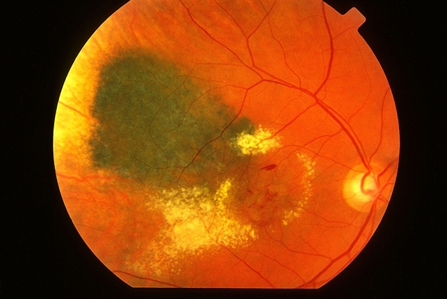
Visual acuity noted was: OD= 6/24, OS= 6/6. On Fundus examination, macular edema, cotton wool spots, and retinal hemorrhages were noted.
Based on history taking and fundus findings, the patient was diagnosed with radiation retinopathy and was given intravitreal bevacizumab as an option of treatment and kept on monthly follow-up.
Visual ups and downs were noted in his follow-up duration and administration of multiple intravitreal injections was made sure from time to time to notice any improvement in visual acuity.
DISEASE
Radiation Retinopathy is a complicated condition in which the retina is damaged due to exposure to any source of radiation used in the treatment of ocular, orbital, or head and neck cancers.
This source of radiation can be either an external beam(which is used in the treatment of nasopharyngeal, paranasal sinuses, and orbital tumors) or plaque brachytherapy (which is used in the treatment of Intraocular tumors).
It was first discovered by Stallard in 1933 and it does not have an acute onset, instead affects after months or years of radiation exposure.
People who are diabetic, hypertensive, those undergoing chemotherapy, and pregnant women are at increased risk of radiation retinopathy.
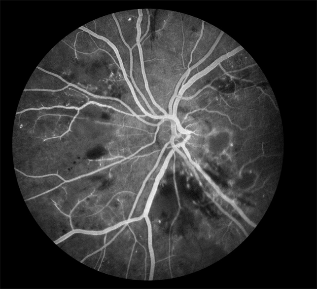
In the early stage of radiation retinopathy, most people are asymptomatic but as the disease progresses, patients may complain of decreased vision and floaters.
Its clinical manifestations are documented as microaneurysms, retinal hemorrhages, cotton wool spots, macular edema, telangiectases, disc edema, perivascular sheathing, capillary non-perfusion, retinal ischemia leading to neovascularization of retina, disc, and iris.
Fundus Explorer Pro
Photograph the retinal findings described here with the phone already in your pocket — 22 D optics and built-in illumination in one handheld unit.
From Choroida — the team behind this siteMANAGEMENT
The management plan includes primary prevention of ocular structures by shielding them during exposure to external beam radiation.
When applying plaque brachytherapy, efforts are made to reduce the dose of radiation by either use of collimating plaques or adjusting the position of plaques eccentrically.
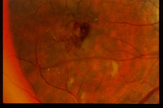
Treatment :
Medical Treatment :
- Intravitreal anti-VEGF agents and corticosteroids are now used to treat radiation retinopathy:
* Intravitreal Bevacizumab temporarily stops the progression of radiation retinopathy. But for long-lasting effects administration of multiple injections is required. It does not improve vision significantly but can prevent further worsening of vision.
* Intravitreal Triamcinolone acetonide is given to treat macular edema.
- Focal laser therapy is done to treat macular edema
- Panretinal photocoagulation is also done to treat regions of ischemia and neovascularization.
Surgical:
Pars plana Vitrectomy(PPV) is done in case of proliferative radiation retinopathy associated with vitreous hemorrhage and tractional retinal detachment.


Document what you see
Two smartphone imaging tools built for everyday clinic use — one for the slit lamp, one for the fundus.
From Choroida — the team behind this siteREFERENCES
- Finger PT, Chin KJ, Duvall G, Palladium-103 for Choroidal Melanoma Study Group. Palladium-103 ophthalmic plaque radiation therapy for choroidal melanoma: 400 treated patients. Ophthalmology. 2009;116(4):790–6. e1. doi: 10.1016/j.ophtha.2008.12.027.
- Wara WM, Irvine AR, Neger RE, Howes EL, Jr, Phillips TL. Radiation retinopathy. Int J Radiat Oncol Biol Phys. 1979 Jan;5(1):81–83.
- Finger PT. Radiation therapy for orbital tumors: concepts, current use, and ophthalmic radiation side effects. Surv Ophthalmol. 2009;54(5):545–568.
- Hayreh SS. Post-radiation retinopathy. A fluorescence fundus angiographic study. Br J Ophthalmol. 1970 Nov;54(11):705–714.
- Noble KG, Kupersmith MJ. Retinal vascular remodelling in radiation retinopathy. Br J Ophthalmol. 1984 Jul;68(7):475–478.
- Gass JD. A fluorescein angiographic study of macular dysfunction secondary to retinal vascular disease. VI. X-ray irradiation, carotid artery occlusion, collagen vascular disease, and vitritis. Arch Ophthalmol. 1968 Nov;80(5):606–617.
Test yourself
A few questions straight from this article.
-
Which two sources of therapeutic radiation can cause radiation retinopathy?
External beam radiation (used for nasopharyngeal, paranasal sinus and orbital tumours) and plaque brachytherapy (used for intraocular tumours) can both damage the retina. -
When does radiation retinopathy typically appear after radiation exposure?
Radiation retinopathy does not have an acute onset; it develops months or years after radiation exposure. -
Which patients are at increased risk of radiation retinopathy?
Diabetes, hypertension, concurrent chemotherapy and pregnancy all increase the risk of radiation retinopathy. -
In radiation retinopathy, what does capillary non-perfusion with retinal ischaemia lead to?
Its manifestations include capillary non-perfusion and retinal ischaemia, which lead to neovascularisation of the retina, disc and iris. -
How is radiation retinopathy prevented during external beam radiation?
Management begins with primary prevention: shielding ocular structures during exposure to external beam radiation. -
How is the retinal radiation dose reduced during plaque brachytherapy to limit radiation retinopathy?
The dose is reduced either by using collimating plaques or by positioning the plaque eccentrically. -
What does intravitreal bevacizumab achieve in radiation retinopathy?
Bevacizumab temporarily stops progression and needs multiple injections for a lasting effect; it prevents further worsening rather than significantly improving vision. -
Which treatments does the article list for macular oedema in radiation retinopathy?
Intravitreal triamcinolone acetonide and focal laser therapy are both used to treat the macular oedema of radiation retinopathy. -
What is panretinal photocoagulation used for in radiation retinopathy?
Panretinal photocoagulation is used to treat regions of retinal ischaemia and neovascularisation. -
When is pars plana vitrectomy indicated in radiation retinopathy?
PPV is performed for proliferative radiation retinopathy associated with vitreous haemorrhage and tractional retinal detachment.
