
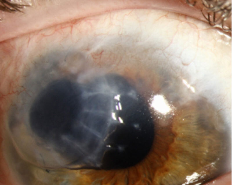
Terrien Marginal Degeneration is a quiet thinning at the edge of the cornea that is easily confused with a far more aggressive disease.
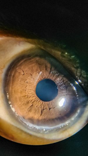
The peripheral cornea slowly melts away, superiorly and painlessly, with a fine line of lipid marking the advancing edge.
Because the epithelium stays intact and the eye is white, it behaves nothing like the painful ulcer it is often mistaken for.
For clinicians, recognizing Terrien Marginal Degeneration matters because distinguishing it from Mooren ulcer and from inflammatory peripheral ulcerative keratitis changes both the work-up and the treatment.
A painless, epithelialised peripheral thinning with lipid is the pattern that names it.
What Is Terrien Marginal Degeneration?
Terrien Marginal Degeneration is a slowly progressive, usually non-inflammatory thinning of the peripheral cornea.
Its defining features are:
- Peripheral corneal thinning, classically superior or superonasal
- An intact epithelium over the thinned area
- A fine lipid line at the leading edge, with superficial vascularization
- A white, painless eye in the typical form
It is a degeneration, not an ulcer, and that distinction drives everything that follows.
Epidemiology
The typical patient is fairly characteristic.
- More common in men than women
- Usually presents in middle age, though a younger inflammatory variant exists
- Often bilateral, but frequently asymmetric
The slow, bilateral, painless course helps separate it from acute inflammatory peripheral disease.
All-fit Slit-Lamp Adapter
Record and share exactly what you see at the slit lamp. One adapter fits any slit lamp or surgical microscope — and any smartphone.
From Choroida — the team behind this siteClinical Presentation
Symptoms
- Often asymptomatic in the early stages
- Gradually blurred vision from induced astigmatism
- Rarely, sudden pain and visual loss if the thinned cornea perforates after minor trauma
Examination Findings
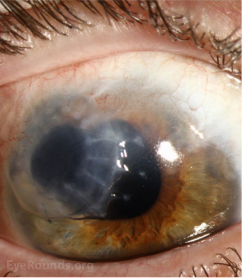
- A gutter of peripheral thinning with an intact epithelium
- A yellow-white lipid deposit at the central edge of the thinning
- Fine superficial vascularization crossing into the gutter
- Against-the-rule or oblique astigmatism on topography
Peripheral thinning with lipid and pannus, but no epithelial defect and no pain, is the fundus of the diagnosis.
Terrien Versus Its Mimics
The differential is the whole clinical point.
- Mooren ulcer — painful, with an epithelial defect and an overhanging central edge that progresses circumferentially and centrally
- Peripheral ulcerative keratitis from rheumatoid arthritis and vasculitis — inflamed, painful, and systemically driven
- Pellucid marginal degeneration — inferior thinning without lipid or vascularization
- Furrow degeneration — a benign thinning in the arcus region without progression
A painless, epithelialised, lipid-lined gutter separates Terrien from the painful, ulcerating mimics that need urgent systemic work-up.
Diagnostic Evaluation
The assessment confirms the pattern and excludes inflammation.
- Slit-lamp examination documents the thinning, lipid, and intact epithelium
- Corneal topography maps the induced astigmatism
- Anterior segment OCT quantifies the residual corneal thickness
- Systemic inflammatory work-up when the presentation is atypical or painful
Documenting an intact epithelium is what keeps the diagnosis in the degenerative rather than the ulcerative camp.
Management
Most cases need refraction, not surgery.
- Correction of astigmatism with spectacles or rigid contact lenses for the common, slowly progressive form
- Tectonic grafting — a patch or crescentic graft — for severe thinning or perforation risk
- Protective eyewear to reduce the risk of traumatic perforation
- Anti-inflammatory treatment for the rarer inflammatory variant
The aim is to manage the astigmatism and protect a thinned cornea, intervening surgically only when perforation threatens.
Prognosis
The outlook is generally good.
- Vision is usually maintained with refractive correction
- Progression is slow in the typical form
- Perforation is uncommon but is the main serious risk
In practice, the value of naming Terrien Marginal Degeneration is sparing the patient the aggressive treatment that Mooren ulcer would demand.
Would you like to document peripheral corneal thinning with your smartphone?


Document what you see
Two smartphone imaging tools built for everyday clinic use — one for the slit lamp, one for the fundus.
From Choroida — the team behind this siteReferences
- Austin P, Brown SI. “Inflammatory Terrien’s Marginal Corneal Disease.” American Journal of Ophthalmology. 1981;92(2):189–192.
- Chan AT, Ulate R, Goldich Y, et al. “Terrien Marginal Degeneration: Clinical Characteristics and Outcomes.” American Journal of Ophthalmology. 2015;160(5):867–872.
- Ding Y, Murri MS, Birdsong OC, et al. “Terrien Marginal Degeneration.” Survey of Ophthalmology. 2019;64(2):162–174.
- American Academy of Ophthalmology. “Terrien Marginal Degeneration.” EyeWiki.
Test yourself
A few questions straight from this article.
-
In Terrien marginal degeneration, where is the peripheral corneal thinning classically located?
The thinning gutter of Terrien marginal degeneration characteristically begins superiorly or superonasally, unlike the inferior thinning of pellucid marginal degeneration. -
Which examination feature keeps Terrien marginal degeneration in the degenerative rather than ulcerative category?
Documenting an intact epithelium over the thinned area is what separates a degeneration from an ulcerating peripheral keratitis. -
What marks the leading central edge of the thinning gutter in Terrien marginal degeneration?
A fine yellow-white lipid line at the advancing central edge, with superficial vessels crossing into the gutter, is the signature of Terrien. -
Which patient profile is typical of Terrien marginal degeneration?
Terrien is commoner in men, usually presents in middle age and is often bilateral though asymmetric; a younger inflammatory variant also exists. -
Which feature points to Mooren ulcer rather than Terrien marginal degeneration?
Mooren ulcer is painful, breaks the epithelium and has an overhanging central edge that advances circumferentially and centrally. -
Which peripheral corneal disorder causes inferior thinning without lipid or vascularization?
Pellucid marginal degeneration thins the inferior cornea and lacks both the lipid line and the superficial vascularization seen in Terrien. -
What astigmatism pattern does corneal topography typically show in Terrien marginal degeneration?
Superior thinning flattens the vertical meridian, so topography maps against-the-rule or oblique astigmatism, which explains the gradual blurring. -
Which investigation quantifies the residual corneal thickness in Terrien marginal degeneration?
Anterior segment OCT measures how much stroma remains in the gutter, which guides whether tectonic support is needed. -
What is the usual first-line management of typical Terrien marginal degeneration?
Most eyes need refraction rather than surgery; spectacles or rigid contact lenses handle the induced astigmatism in the slowly progressive form. -
Which complication is the main serious risk in Terrien marginal degeneration?
Perforation is uncommon but is the main serious risk, which is why protective eyewear and tectonic grafting are used when thinning is severe.
