
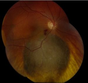
A pigmented spot in the choroid is one of the commonest incidental findings in the fundus, and one of the most anxiety-provoking.
Most are benign choroidal nevi that will never cause harm, yet a small number are early melanomas that can metastasize and kill.
The whole clinical skill lies in telling a harmless choroidal nevus from a small melanoma before it grows.
Fortunately, the decision is not a guess: a set of validated risk factors, remembered by the mnemonic TFSOM, turns a vague worry into a structured judgement.
Knowing these features tells you which lesions can be photographed and watched, and which need urgent referral to an ocular oncologist.
What Is a Choroidal Nevus?
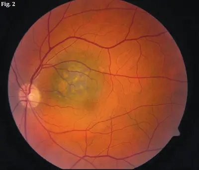
A choroidal nevus is a benign proliferation of melanocytes in the choroid.
Typical, reassuring features are:
- A flat or minimally elevated, slate-grey lesion
- Thickness of 2 mm or less
- Overlying drusen and a surrounding halo
- No subretinal fluid, orange pigment, or symptoms
Drusen and a halo are signs of chronicity and strongly favour a benign nevus.
What Is a Small Choroidal Melanoma?
A small choroidal melanoma is a malignant melanocytic tumour, often indistinguishable from a nevus at a glance.
Worrying features include:
- Increasing thickness, usually beyond 2 mm
- Subretinal fluid and visual symptoms
- Orange pigment (lipofuscin) over the lesion
- A margin touching or near the optic disc
- Documented growth over time
The more of these features a lesion shows, the more likely it is to be a melanoma rather than a nevus.
Fundus Explorer Pro
Photograph the retinal findings described here with the phone already in your pocket — 22 D optics and built-in illumination in one handheld unit.
From Choroida — the team behind this siteThe TFSOM Risk Factors
The risk factors for growth of a small melanocytic lesion are captured by the mnemonic TFSOM-UHHD: “To Find Small Ocular Melanoma – Using Helpful Hints Daily.”
| Letter | Risk factor |
|---|---|
| T | Thickness greater than 2 mm |
| F | Subretinal Fluid |
| S | Symptoms (vision loss) |
| O | Orange pigment |
| M | Margin within 3 mm of the optic disc |
| UH | Ultrasonographic Hollowness |
| H | absence of Halo |
| D | absence of Drusen |
Risk rises with the number of factors: a lesion with none behaves like a nevus, whereas three or more features carry a substantial risk of growth into melanoma.
Diagnostic Evaluation
Multimodal imaging documents the lesion and detects the risk factors objectively.
Ultrasonography
- B-scan measures thickness and detects acoustic hollowness
- Increasing thickness on serial scans is a powerful sign of malignancy
OCT and Autofluorescence
- OCT is the most sensitive way to detect subretinal fluid
- Fundus autofluorescence highlights orange pigment as bright hyper-autofluorescence
Photographic Surveillance
- Widefield colour photography documents the margins and size for comparison
- Serial images are the simplest way to prove or exclude growth
Objective imaging turns a subjective impression into a documented, comparable record.
Differential Diagnosis
Other pigmented or elevated choroidal lesions can enter the differential:
- Choroidal melanocytoma — typically jet-black, often juxtapapillary
- Congenital hypertrophy of the RPE — flat, well-defined, with lacunae
- Choroidal metastasis — often creamy and multifocal
- Peripheral exudative haemorrhagic chorioretinopathy — a haemorrhagic peripheral mimic
- Congenital or reactive RPE hyperplasia
Applying the TFSOM factors alongside this differential keeps both over- and under-diagnosis in check.
Management
Management follows directly from the risk assessment.
- Low-risk nevi (no risk factors) are photographed and reviewed periodically
- Intermediate lesions warrant closer follow-up with imaging
- High-risk lesions (three or more factors, or documented growth) need prompt referral to an ocular oncology service
- Confirmed small melanomas are treated with plaque brachytherapy or other globe-conserving therapy
The safest rule is to refer any lesion whose features or growth raise genuine concern.
Prognosis
Outcomes depend heavily on early recognition.
- The vast majority of choroidal nevi remain stable for life
- Small melanomas detected and treated early have a far better prognosis than thick tumours
- Delay allows growth, metastatic risk, and loss of the eye
In practice, the value of the TFSOM factors is catching the rare dangerous lesion while it is still small and treatable.


Document what you see
Two smartphone imaging tools built for everyday clinic use — one for the slit lamp, one for the fundus.
From Choroida — the team behind this siteReferences
- Shields CL, Shields JA, Kiratli H, et al. “Risk Factors for Growth and Metastasis of Small Choroidal Melanocytic Lesions.” Ophthalmology. 1995;102(9):1351–1361.
- Shields CL, Furuta M, Berman EL, et al. “Choroidal Nevus Transformation Into Melanoma: Analysis of 2514 Consecutive Cases.” Archives of Ophthalmology. 2009;127(8):981–987.
- Singh AD, Kalyani P, Topham A. “Estimating the Risk of Malignant Transformation of a Choroidal Nevus.” Ophthalmology. 2005;112(10):1784–1789.
- Shields CL, Dalvin LA, Yu MD, et al. “Choroidal Nevus Transformation Into Melanoma Per Millimeter Increment in Thickness Using Multimodal Imaging.” Retina. 2019;39(10):1852–1860.
- American Academy of Ophthalmology. “Choroidal Nevus.” EyeWiki.
Test yourself
A few questions straight from this article.
-
A choroidal nevus is best described as which type of lesion?
A choroidal nevus is a benign melanocytic proliferation, classically a flat or minimally elevated slate-grey lesion 2 mm thick or less. -
Which pair of findings over a choroidal melanocytic lesion is most reassuring for a benign nevus?
Drusen and a halo are signs of chronicity and strongly favour a benign nevus; their absence is itself counted among the TFSOM risk factors. -
In the TFSOM risk factors, what thickness of a choroidal melanocytic lesion counts as high risk?
The T in TFSOM stands for thickness greater than 2 mm; a typical benign nevus measures 2 mm or less. -
Which surface pigment over a choroidal lesion is a TFSOM risk factor for melanoma?
Orange pigment, which is lipofuscin, is the O of TFSOM and appears as bright hyper-autofluorescence on fundus autofluorescence. -
How close to the optic disc must a lesion margin be to count as a TFSOM risk factor?
The M of TFSOM refers to a lesion margin lying within 3 mm of the optic disc. -
Which ultrasonographic feature is included among the TFSOM risk factors for choroidal melanoma?
Ultrasonographic hollowness is the UH of TFSOM-UHHD, and serial B-scan also measures thickness, where increasing values strongly suggest malignancy. -
Which imaging test is the most sensitive way to detect subretinal fluid over a choroidal nevus?
OCT detects subretinal fluid more sensitively than any other modality, and subretinal fluid is the F of the TFSOM risk factors. -
What is the simplest way to prove or exclude growth of a choroidal melanocytic lesion over time?
Widefield colour photography documents lesion margins and size, and serial images provide the simplest objective comparison for growth. -
How should a choroidal melanocytic lesion with three or more TFSOM risk factors be managed?
Three or more risk factors, or documented growth, mark a high-risk lesion carrying a substantial risk of growth into melanoma and require prompt ocular oncology referral. -
Which treatment is used for a confirmed small choroidal melanoma?
Confirmed small melanomas are treated with plaque brachytherapy or another globe-conserving therapy, and early treatment carries a far better prognosis than thick tumours.

Writes and reviews the clinical articles on Educate. Every post is checked against peer-reviewed sources before it goes live.


