
Pars planitis is a chronic idiopathic form of intermediate uveitis, characterized by vitritis, snowballs, and a snowbank of exudate over the inferior pars plana.
It typically affects children, adolescents, and young adults, and it often presents with floaters and blurred vision in an eye that looks white and comfortable.
Macular edema is the main threat to vision, and the disease can smolder for years before it is recognized.
What Is Pars Planitis?
The Standardization of Uveitis Nomenclature (SUN) working group defines intermediate uveitis as inflammation that is primarily in the vitreous, with or without peripheral retinal vascular sheathing.
Pars planitis is the subset with snowbank or snowball formation and no associated infection or systemic disease.
When intermediate uveitis is linked to a specific condition, such as multiple sclerosis or sarcoidosis, the term used is intermediate uveitis associated with that disease.
The disease is typically bilateral, although it is often asymmetric, and it accounts for a moderate proportion of uveitis in children.
Clinical Features
Symptoms
Floaters and blurred vision are the most common complaints.
Pain, redness, and photophobia are usually absent, which is why the disease may be discovered late.
Signs
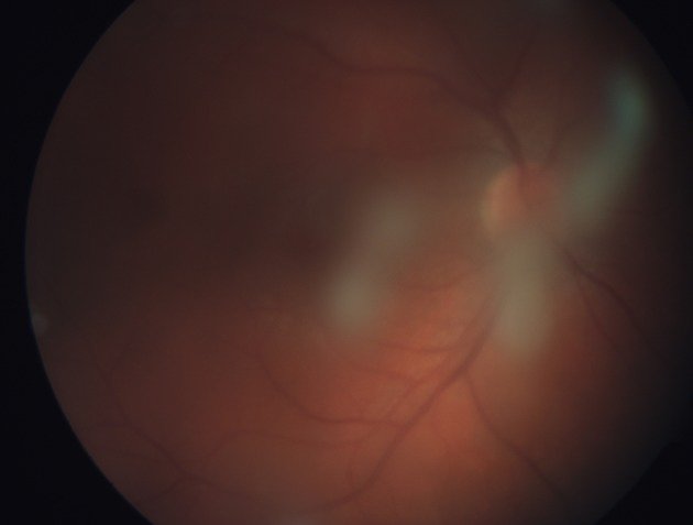
- Vitreous cells and haze, often the dominant finding
- Snowballs, which are aggregates of inflammatory cells in the inferior vitreous
- Snowbanking, a white fibrovascular exudate over the inferior pars plana and peripheral retina
- Peripheral retinal periphlebitis, seen as sheathing and leakage
- Mild anterior chamber cells and, in some patients, band keratopathy in children
- Macular edema, which may be cystoid or diffuse
Complications
- Cystoid macular edema, the leading cause of vision loss
- Posterior subcapsular cataract, from disease or steroid treatment
- Glaucoma from inflammation and steroids
- Epiretinal membrane and vitreous hemorrhage
- Peripheral neovascularization at the snowbank, sometimes with vitreous hemorrhage
- Tractional or rhegmatogenous detachment in advanced disease
In a 20-year population study from the United States, macular edema and cataract were the most common complications, and vision was good in most patients at last follow-up.
Fundus Explorer Pro
Photograph the retinal findings described here with the phone already in your pocket — 22 D optics and built-in illumination in one handheld unit.
From Choroida — the team behind this sitePars Planitis in Children
Children with pars planitis often present late because they do not complain of floaters and their eyes are white.
The condition may be found on routine screening, or when a child develops squint, poor school performance, or leukocoria-like cataract.
Band keratopathy, posterior subcapsular cataract, and macular edema are frequent at diagnosis.
The disease can be more aggressive in young children, and the risk of amblyopia adds to the visual burden.
Examination under anesthesia may be needed for a complete peripheral assessment, and long-term steroid use has to be limited because of growth and cataract risks.
Differential Diagnosis and Workup
Pars planitis is a diagnosis of exclusion.
The workup aims to identify treatable causes and associated systemic disease.
- Syphilis serology, since syphilis mimics nearly every form of uveitis (see ocular syphilis)
- Tuberculosis testing and chest imaging (see ocular tuberculosis)
- Chest imaging and serum ACE for sarcoidosis (see ocular sarcoidosis)
- MRI of the brain when there are neurologic symptoms or demyelination is a concern
- Lyme serology in endemic areas
- Vitreous sampling when lymphoma or another masquerade is possible, particularly in older adults
Infectious and inflammatory mimics that resemble pars planitis include toxocariasis in children, cat scratch disease, and intraocular lymphoma.
Multiple sclerosis is reported in a proportion of patients with intermediate uveitis, so neurologic symptoms should be sought and MRI considered.
Investigations
OCT quantifies macular edema and shows epiretinal membranes.
Fluorescein angiography shows peripheral vascular leakage and disc hyperfluorescence, and widefield angiography helps identify neovascularization at the snowbank.
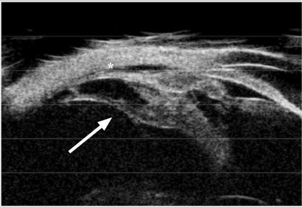
B-scan or ultrasound biomicroscopy is helpful when the view is poor and shows pars plana exudates.
Management
Treatment is not always needed.
Patients with good vision and no macular edema can be observed.
Treatment is started for vision-threatening complications, particularly macular edema, and should follow a stepwise plan.
Local and Systemic Corticosteroids
- Periocular or intravitreal triamcinolone for unilateral disease
- Dexamethasone or fluocinolone implants for persistent or recurrent disease
- Oral prednisone for bilateral disease with macular edema
Steroid side effects include cataract and raised IOP, and children need close monitoring because of growth and glaucoma risk.
Immunomodulatory Therapy
Steroid-sparing therapy is used when disease is bilateral, chronic, or steroid-dependent.
Options include methotrexate, mycophenolate, azathioprine, cyclosporine, and biologics such as adalimumab.
Retinal Ablation
Cryotherapy or laser to the snowbank is used for peripheral neovascularization, and the aim is to regress the new vessels (see retinal cryotherapy).
Surgery
Pars plana vitrectomy is useful for nonclearing vitreous hemorrhage, tractional detachment, epiretinal membranes, and refractory macular edema (see pars plana vitrectomy).
Cataract surgery should be done when inflammation has been controlled for several months, with perioperative steroid cover.
Follow-Up
Patients on treatment or with active disease are reviewed every one to three months at first, with OCT at each visit.
After inflammation is quiet for a prolonged period, intervals can lengthen to every six months.
Each visit should include IOP measurement, a check for cataract progression, and a dilated peripheral examination for new snowbank activity or neovascularization.
Patients should be told to return promptly for new floaters, a sudden increase in haze, or a curtain, because retinal tears and detachment can complicate long-standing intermediate uveitis.
Prognosis
The course is variable.
Some patients have a self-limited course over years, others have persistent inflammation with recurrent macular edema.
Long-term visual outcome is good in most patients who are followed and treated appropriately.
Because the disease is chronic and complications accumulate slowly, regular OCT and IOP monitoring are essential.


Document what you see
Two smartphone imaging tools built for everyday clinic use — one for the slit lamp, one for the fundus.
From Choroida — the team behind this siteReferences
- Jabs DA, Nussenblatt RB, Rosenbaum JT; Standardization of Uveitis Nomenclature (SUN) Working Group. Standardization of uveitis nomenclature for reporting clinical data. Results of the First International Workshop. Am J Ophthalmol. 2005;140:509-516.
- Donaldson MJ, Pulido JS, Herman DC, Diehl N, Hodge D. Pars planitis: a 20-year study of incidence, clinical features, and outcomes. Am J Ophthalmol. 2007;144:812-817.
- Malinowski SM, Pulido JS, Folk JC. Long-term visual outcome and complications associated with pars planitis. Ophthalmology. 1993;100:818-824.
- Bonfioli AA, Orefice F. Intermediate uveitis. Semin Ophthalmol. 2005;20:147-154.
- Kempen JH, Altaweel MM, Holbrook JT, et al. Randomized comparison of systemic anti-inflammatory therapy versus fluocinolone acetonide implant for intermediate, posterior, and panuveitis: the Multicenter Uveitis Steroid Treatment Trial. Ophthalmology. 2011;118:1916-1926.
