
Goniotomy is an angle surgery that directly incises the abnormal or obstructed trabecular meshwork under gonioscopic visualization, remaining the primary surgical treatment for primary congenital glaucoma, discussed in its own dedicated article on this site.
Unlike trabeculectomy or tube shunt surgery, both discussed in their own dedicated articles on this site, goniotomy works entirely from inside the eye through a clear corneal incision, directly addressing the developmental trabecular meshwork abnormality thought to underlie most cases of primary congenital glaucoma.
Understanding why this direct, ab-interno approach is so effective specifically in congenital glaucoma, and less broadly applicable in adult glaucoma, comes down to the very different underlying mechanisms driving pressure elevation in these two very different patient populations.
Modern angle surgery for adult glaucoma, including several MIGS procedures discussed in their own dedicated article on this site, actually borrows conceptually from this same angle-based approach, even though the specific technique and target patient population differ considerably.

Why It Works So Well in Congenital Glaucoma
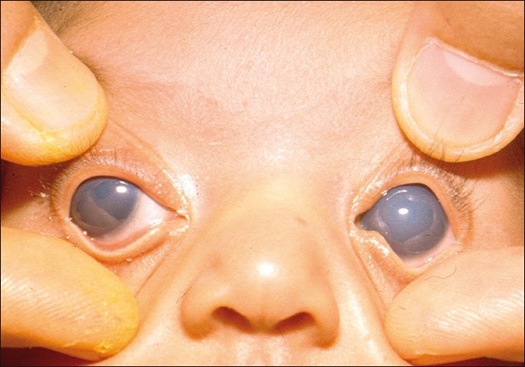
Primary congenital glaucoma is thought to result from abnormal, incompletely developed trabecular meshwork tissue that obstructs normal aqueous outflow, a fundamentally different mechanism from the age-related or otherwise acquired outflow changes seen in adult glaucoma.
Because the obstruction in congenital glaucoma sits directly at the level of this abnormal trabecular tissue, physically incising it removes the obstruction at its actual anatomic source, restoring more direct access to the eye’s normal downstream outflow pathway.
This direct correspondence between the surgical technique and the specific underlying developmental abnormality is why goniotomy achieves such favorable success rates in appropriately selected congenital glaucoma cases, a result not generally replicated when similar angle surgery is attempted in adult open-angle glaucoma.
Technique
The surgeon visualizes the angle directly using a specialized goniolens, discussed in relation to gonioscopy in its own dedicated article on this site, while working through a clear corneal paracentesis incision rather than a scleral approach.
A fine goniotomy knife or needle is passed across the anterior chamber to incise the trabecular meshwork along a portion of the angle’s circumference, creating a direct communication between the anterior chamber and Schlemm’s canal.
Adequate corneal clarity is essential for this technique, since the surgeon depends entirely on a clear view through the goniolens to safely and accurately perform the incision, which is a real practical limitation in eyes with significant corneal edema from very high pressure.
All-fit Slit-Lamp Adapter
Record and share exactly what you see at the slit lamp. One adapter fits any slit lamp or surgical microscope — and any smartphone.
From Choroida — the team behind this siteGoniotomy Versus Trabeculotomy
Trabeculotomy achieves a similar surgical goal, opening the trabecular meshwork and Schlemm’s canal, but approaches from outside the eye through a scleral incision, threading a probe into Schlemm’s canal and rotating it inward to rupture the trabecular meshwork from behind.
Trabeculotomy does not require the same degree of corneal clarity that goniotomy does, since it does not rely on a direct gonioscopic view through the cornea, making it a useful alternative specifically in eyes where corneal edema limits adequate visualization for goniotomy.
Both procedures share the same underlying surgical goal and are used somewhat interchangeably depending on surgeon preference, corneal clarity, and specific anatomic considerations in an individual case.
Newer instrumentation, including illuminated microcatheters that allow a full 360-degree circumferential trabeculotomy rather than a more limited segment, has further blurred the practical distinction between the two techniques in some modern practices.
Outcomes and Repeat Procedures
- Success rates are generally favorable in appropriately selected primary congenital glaucoma, particularly when performed relatively early in the disease course
- Repeat goniotomy, sometimes addressing a different portion of the angle’s circumference than the first procedure, is a reasonable option when an initial treatment does not achieve adequate pressure control
- Eyes that fail one or more angle surgeries may require trabeculectomy or a glaucoma drainage device, following the broader pediatric glaucoma treatment ladder discussed in relation to aphakic glaucoma in its own dedicated article on this site
Postoperative Care
Close monitoring of intraocular pressure, corneal clarity, and axial length or corneal diameter in a young child is essential after goniotomy, mirroring the lifelong surveillance principles discussed for pediatric glaucoma more broadly.
A degree of postoperative hyphema is common and generally self-limited, typically clearing over the days following surgery without specific intervention beyond routine monitoring.
Given the developmental nature of the underlying condition and the possibility of needing further intervention, families are counseled from the outset that ongoing follow-up, and possibly repeat surgery, is a normal, anticipated part of managing this condition rather than a sign that the initial procedure failed.
Examination under anesthesia is often needed at multiple points during a young child’s care, both to perform the initial surgery itself and to obtain reliable pressure measurements and other exam findings that an awake infant or toddler cannot otherwise provide.


Document what you see
Two smartphone imaging tools built for everyday clinic use — one for the slit lamp, one for the fundus.
From Choroida — the team behind this siteReferences
- American Academy of Ophthalmology. Basic and Clinical Science Course, Section 6: Pediatric Ophthalmology and Strabismus.
- Russell-Eggitt IM, Rice NS, Jay B, Wyse RK. Relapse following goniotomy for congenital glaucoma due to trabecular dysgenesis. Eye.
- Girkin CA. Goniotomy versus trabeculotomy. Journal of Glaucoma.
Test yourself
A few questions straight from this article.
-
Goniotomy lowers intraocular pressure by directly incising which structure?
Goniotomy is an angle surgery that incises the abnormal or obstructed trabecular meshwork under gonioscopic visualisation, and it remains the primary surgical treatment for primary congenital glaucoma. -
Pressure elevation in primary congenital glaucoma is thought to arise from which abnormality?
Primary congenital glaucoma is thought to result from abnormal, incompletely developed trabecular meshwork obstructing outflow, a mechanism quite different from the acquired outflow changes of adult glaucoma. -
Through which route does the surgeon reach the angle during goniotomy?
Goniotomy works entirely from inside the eye through a clear corneal paracentesis incision rather than a scleral approach, unlike trabeculectomy or tube shunt surgery. -
Incising the meshwork during goniotomy creates direct communication between which two spaces?
A fine goniotomy knife or needle incises the trabecular meshwork along part of the angle's circumference, creating direct communication between the anterior chamber and Schlemm's canal. -
Which requirement limits goniotomy in an infant whose pressure has been very high?
The surgeon depends entirely on a clear view through the goniolens, so significant corneal edema from very high pressure is a real practical limitation to goniotomy. -
How does trabeculotomy open the trabecular meshwork?
Trabeculotomy approaches from outside the eye through a scleral incision, threading a probe into Schlemm's canal and rotating it inward to rupture the trabecular meshwork from behind. -
Which situation makes trabeculotomy preferable to goniotomy in congenital glaucoma?
Trabeculotomy does not rely on a direct gonioscopic view through the cornea, making it a useful alternative specifically in eyes where corneal edema limits visualisation. -
Illuminated microcatheters have changed trabeculotomy by allowing which modification?
Illuminated microcatheters permit a full 360-degree circumferential trabeculotomy rather than a limited segment, blurring the practical distinction between trabeculotomy and goniotomy. -
When initial goniotomy fails to control pressure in congenital glaucoma, what is a reasonable next step?
Repeat goniotomy, often addressing a different portion of the angle's circumference, is a reasonable option when the first procedure does not achieve adequate pressure control. -
Which postoperative finding after goniotomy is common and usually self-limited?
A degree of postoperative hyphema is common after goniotomy and generally self-limited, clearing over the days after surgery without intervention beyond routine monitoring.

Writes and reviews the clinical articles on Educate. Every post is checked against peer-reviewed sources before it goes live.


