
Von Hippel-Lindau disease is an autosomal dominant tumour suppressor syndrome, and the retinal capillary hemangioblastoma is often the first manifestation to appear clinically, sometimes years before the cerebellar, renal, or adrenal tumours that make this a genuinely dangerous multisystem disease.
That timing matters enormously in practice: an ophthalmologist who recognises a retinal capillary hemangioblastoma and pursues systemic screening can be the reason a patient’s renal cell carcinoma or cerebellar haemangioblastoma is caught years before it would otherwise have presented.
Genetics
Von Hippel-Lindau disease results from a germline mutation in the VHL tumour suppressor gene, with tumour formation following the classic two-hit model, where a second, somatic mutation in the remaining normal allele triggers formation of a tumour at a given site.
Because the germline mutation is present in every cell, patients are at risk of multiple, often multifocal tumours across several organ systems over their lifetime, which is the underlying reason lifelong systemic surveillance, not a one-time evaluation, is central to management.
Retinal Capillary Hemangioblastoma
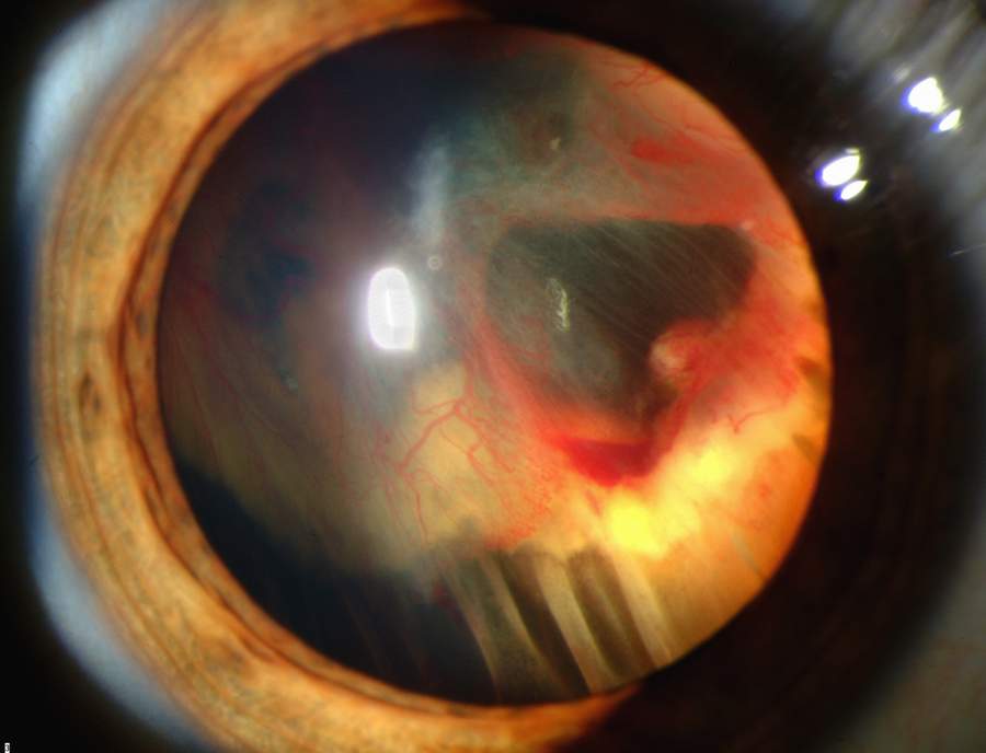
These are benign vascular tumours of the retina, appearing as an orange-red mass with characteristically dilated, tortuous feeder and draining vessels connecting it to the retinal circulation, a pattern that is often visually distinctive enough to suggest the diagnosis on fundus examination alone.
Lesions are frequently multiple and bilateral, and can occur anywhere in the retina, though the periphery is a common location, sometimes making them easy to miss without a careful, complete peripheral examination.
Exudation from the tumour can produce lipid deposition in the macula at a considerable distance from the lesion itself, and larger or more active tumours carry a risk of exudative retinal detachment, vitreous haemorrhage, and secondary glaucoma if left untreated.
Fundus Explorer Pro
Photograph the retinal findings described here with the phone already in your pocket — 22 D optics and built-in illumination in one handheld unit.
From Choroida — the team behind this siteSystemic Manifestations
- Cerebellar and spinal haemangioblastoma, the most common central nervous system tumour in this disease, which can cause headache, ataxia, or other neurological symptoms depending on location
- Renal cell carcinoma, often multiple and bilateral, and a leading cause of mortality in this disease if not identified and managed through regular screening
- Phaeochromocytoma, which can cause hypertension and should be screened for, particularly relevant before any surgical procedure given the anaesthetic risks of an unrecognised phaeochromocytoma
- Pancreatic cysts and neuroendocrine tumours
- Endolymphatic sac tumours, a less common but recognised manifestation that can affect hearing
Diagnostic Evaluation
Diagnosis is established through genetic testing for VHL mutations in a patient with a suggestive clinical picture, or through clinical criteria based on the pattern and number of characteristic tumours in patients with or without a known family history.
Once diagnosed, or in an at-risk relative of a known patient, a structured, lifelong surveillance protocol is followed, including regular dilated fundus examination, MRI of the brain and spine, renal imaging, and biochemical screening for phaeochromocytoma, since the goal is to catch each tumour type while it remains small and manageable rather than after it has caused symptoms.
Differential Diagnosis
- Coats disease, which also causes retinal telangiectasia and exudation but typically presents in a younger, unilateral pattern without the characteristic feeder and draining vessels or the systemic tumour associations of VHL disease
- Retinal cavernous hemangioma, a different vascular malformation with a cluster-of-grapes appearance and generally no feeder vessels, and without the same systemic tumour risk
- Racemose haemangioma (Wyburn-Mason syndrome), an arteriovenous malformation of the retina associated with similar lesions intracranially, distinguished by its different vascular architecture and associations
- Choroidal melanoma or other pigmented or vascular tumours, distinguished on careful examination and imaging by their different appearance and lack of the specific feeder-vessel pattern
Management
Treating the Retinal Tumour
Small, asymptomatic peripheral lesions may be observed with regular monitoring, while laser photocoagulation or cryotherapy is used for small to moderate-sized tumours to control growth and exudation before they threaten vision.
Larger tumours, or those causing significant exudation or detachment, may require more intensive treatment, including photodynamic therapy, anti-VEGF therapy as an adjunct, or, in severe cases with extensive detachment, vitrectomy surgery.
Systemic Screening and Management
Coordinated, lifelong screening for cerebellar, renal, adrenal, and pancreatic manifestations is managed jointly with genetics, neurosurgery, urology, and endocrinology, since each of these tumour types has its own surveillance schedule and treatment threshold, and ophthalmology is only one part of a much larger, structured surveillance programme.
Family Screening
Genetic counselling and testing of at-risk relatives allows early identification of affected family members before symptoms develop, extending the benefit of early detection beyond just the index patient.
Prognosis
Visual outcome from retinal capillary hemangioblastoma is generally good when tumours are identified and treated while still small, though larger or neglected lesions can cause significant, sometimes permanent, vision loss from exudative detachment or macular involvement.
Overall prognosis for the patient is dominated by the systemic manifestations of the disease, particularly renal cell carcinoma and central nervous system haemangioblastoma, which is why structured lifelong surveillance across multiple organ systems, not just periodic eye examinations, is what actually protects these patients’ long-term health.
Early detection through screening, whether prompted by an ophthalmologist finding a retinal lesion or by systematic family screening once one member is diagnosed, is consistently associated with better outcomes across every organ system this disease can affect, which is the central argument for the surveillance-based approach that defines modern management of VHL disease.


Document what you see
Two smartphone imaging tools built for everyday clinic use — one for the slit lamp, one for the fundus.
From Choroida — the team behind this siteReferences
- Maher ER, Neumann HP, Richard S. von Hippel-Lindau disease: a clinical and scientific review. European Journal of Human Genetics. 2011.
- Singh AD, Nouri M, Shields CL, et al. Treatment of retinal capillary hemangioma. Ophthalmology. 2002.
- Chew EY. Ocular manifestations of von Hippel-Lindau disease: clinical and genetic investigations. Transactions of the American Ophthalmological Society. 2005.
- Von Hippel-Lindau Disease. EyeWiki, American Academy of Ophthalmology.
- Von Hippel-Lindau Syndrome. StatPearls, NCBI Bookshelf.
Test yourself
A few questions straight from this article.
-
What is the genetic basis of von Hippel-Lindau disease?
This autosomal dominant tumour suppressor syndrome follows the two-hit model: a germline VHL mutation plus a somatic mutation in the remaining allele triggers a tumour at a given site. -
What is the characteristic fundus appearance of a retinal capillary hemangioblastoma?
The orange-red vascular tumour with characteristically dilated, tortuous feeder and draining vessels is often distinctive enough to suggest the diagnosis on fundus examination alone. -
Where are retinal capillary hemangioblastomas typically found in von Hippel-Lindau disease?
Lesions are commonly multiple and bilateral and can occur anywhere in the retina, with the periphery a frequent site, so they are easily missed without a complete peripheral examination. -
Why can a peripheral retinal capillary hemangioblastoma reduce central vision?
Exudation from the tumour can lay down lipid in the macula well away from the lesion itself, so a peripheral tumour can threaten central acuity. -
Which complications threaten the eye from an untreated retinal capillary hemangioblastoma?
Larger or more active tumours risk exudative retinal detachment, vitreous haemorrhage and secondary glaucoma if they are left untreated. -
Which tumour is a leading cause of mortality in von Hippel-Lindau disease?
Renal cell carcinoma, often multiple and bilateral, is a leading cause of death in this disease when it is not identified and managed through regular screening. -
What does lifelong surveillance in von Hippel-Lindau disease involve?
Structured surveillance combines regular dilated fundus examination, MRI of brain and spine, renal imaging and biochemical phaeochromocytoma screening, aiming to catch each tumour while still small. -
Why does phaeochromocytoma screening matter before surgery in von Hippel-Lindau disease?
Phaeochromocytoma can cause hypertension and should be screened for, particularly before any surgical procedure, given the anaesthetic risks if it goes unrecognised. -
What distinguishes Coats disease from a retinal capillary hemangioblastoma?
Coats disease also causes retinal telangiectasia and exudation, but typically presents unilaterally in a younger patient, without the feeder and draining vessels or systemic tumour associations of VHL. -
How are small to moderate-sized retinal capillary hemangioblastomas usually treated?
Laser photocoagulation or cryotherapy is used to control growth and exudation before the tumour threatens vision, while small asymptomatic peripheral lesions may simply be monitored.

Writes and reviews the clinical articles on Educate. Every post is checked against peer-reviewed sources before it goes live.


