
Lateral canthotomy and cantholysis is an emergency bedside procedure to relieve orbital compartment syndrome, most often from retrobulbar haemorrhage, by releasing the lateral canthal tendon and creating space for orbital pressure to decompress.
It is one of the few genuinely time-critical procedures in ophthalmology. Retinal and optic nerve ischaemia can begin within roughly 90 to 120 minutes of significantly elevated orbital pressure, and permanent vision loss follows if the pressure is not relieved.
The procedure does not require an operating theatre, general anaesthesia, or specialised equipment, which is precisely the point: it needs to be performed wherever the patient is found to be at risk, often in an emergency department by whoever is present and trained to do it.
Recognising Orbital Compartment Syndrome
The orbit is a closed bony compartment, and any process that adds volume within it, most commonly haemorrhage after trauma or surgery, but also severe orbital cellulitis or emphysema, raises orbital pressure with nowhere for that pressure to escape.
As pressure rises, it compresses the central retinal artery and optic nerve, threatening irreversible ischaemic damage if not relieved promptly.
Clinical signs include proptosis, tense resistance to retropulsion of the globe, marked reduction in visual acuity, a relative afferent pupillary defect, elevated intraocular pressure, and pain out of proportion to the apparent injury. Not every sign needs to be present to justify urgent action; a marked drop in vision with a tense orbit after trauma is enough to proceed.
Indications for the Procedure
- Clinical diagnosis of orbital compartment syndrome with signs of optic nerve or retinal compromise, such as reduced vision, a relative afferent pupillary defect, or markedly elevated intraocular pressure
- A tense, non-compressible orbit on examination following trauma, surgery, or rapidly progressive orbital swelling
- The decision to proceed should be based on clinical findings, not on waiting for imaging confirmation, since the time pressure of ischaemic injury does not allow for a CT scan to be completed first when signs are already present
All-fit Slit-Lamp Adapter
Record and share exactly what you see at the slit lamp. One adapter fits any slit lamp or surgical microscope — and any smartphone.
From Choroida — the team behind this siteProcedure
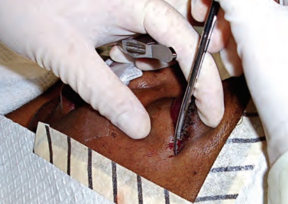
Local anaesthetic is infiltrated at the lateral canthus if time allows, though the procedure should not be delayed for anaesthesia in a genuine emergency with vision already threatened.
A haemostat is used to crush the lateral canthal skin for a few seconds, which reduces bleeding at the incision site. Scissors are then used to cut horizontally through the full thickness of the lateral canthus, performing the canthotomy.
The lower eyelid is then grasped and pulled away from the globe to place the inferior crus of the lateral canthal tendon under tension, which is identified and cut, releasing the lower lid from its lateral attachment. This step, cantholysis, is what actually decompresses the orbit; the canthotomy alone is not sufficient.
If pressure remains elevated and vision does not improve after inferior cantholysis, the superior crus can also be released for further decompression.
Confirming Success
Improvement in visual acuity, softening of the globe on palpation, and reduction in intraocular pressure are the immediate signs that decompression has been effective.
The lower lid should now move freely away from the globe, no longer tethered by the released tendon. If these signs are not seen, reassessing whether a complete cantholysis was achieved, and considering release of the superior crus, is the next step.
Differential Diagnosis
Other causes of acute proptosis and orbital pain need to be considered, though most do not present with the same degree of tension and rapid visual compromise that mandates emergency canthotomy.
- Orbital cellulitis, which can also raise orbital pressure but typically evolves over hours to days rather than the acute onset of traumatic haemorrhage
- Carotid-cavernous fistula, causing chronic pulsatile proptosis rather than the acute, rapidly progressive picture of compartment syndrome
- Orbital emphysema following facial fracture, which can occasionally raise pressure enough to threaten the optic nerve, particularly after forceful nose-blowing, and is managed with the same decompression approach if compartment syndrome develops
Aftercare
Once emergency decompression has been performed, urgent ophthalmology involvement is needed for further management, definitive control of the underlying cause of bleeding or swelling, and formal closure or repair of the canthal defect once the acute crisis has passed.
Imaging, typically CT of the orbits, is obtained once the patient is stabilised to characterise the extent of haemorrhage or other pathology and guide further management, including whether any additional surgical intervention is needed.
Prognosis
Vision recovery after timely canthotomy and cantholysis is generally good when the procedure is performed before irreversible ischaemic damage has occurred, which is why speed matters more than precision in the initial decompression.
Delayed recognition or delayed intervention, waiting for imaging confirmation or specialist availability when signs of compartment syndrome are already present, is the main preventable cause of permanent vision loss in this condition.
The canthal defect created by the procedure heals well with formal repair once the emergency has passed, and cosmetic outcome is rarely a significant long-term concern compared with the vision that was preserved by acting promptly.


Document what you see
Two smartphone imaging tools built for everyday clinic use — one for the slit lamp, one for the fundus.
From Choroida — the team behind this siteReferences
- Lima V, Burt B, Leibovitch I, et al. Orbital compartment syndrome: the ophthalmic surgical emergency. Survey of Ophthalmology. 2009.
- Ballard SR, Enzenauer RW, O’Donnell T, et al. Emergency lateral canthotomy and cantholysis: a simple procedure to preserve vision from sight threatening orbital hemorrhage. Journal of Special Operations Medicine. 2009.
- Vassallo S, Hartstein M, Howard D, Stetz J. Traumatic retrobulbar hemorrhage: emergent decompression by lateral canthotomy and cantholysis. Journal of Emergency Medicine. 2002.
- Lateral Canthotomy and Cantholysis. EyeWiki, American Academy of Ophthalmology.
- Canthotomy and Cantholysis. StatPearls, NCBI Bookshelf.
Test yourself
A few questions straight from this article.
-
What is the purpose of emergency lateral canthotomy and cantholysis?
It relieves orbital compartment syndrome, most often from retrobulbar haemorrhage. No theatre, general anaesthesia or special equipment is needed, which is what makes it a bedside procedure. -
Why does orbital haemorrhage raise pressure so dangerously?
Anything adding volume, whether blood, infection or emphysema, raises pressure with nowhere to escape, and that pressure compresses the central retinal artery and optic nerve. -
Within roughly what time can ischaemic damage begin in orbital compartment syndrome?
Retinal and optic nerve ischaemia can start within this window of significantly raised orbital pressure, and permanent visual loss follows if pressure is not relieved. -
Which examination finding supports a diagnosis of orbital compartment syndrome?
Proptosis, a relative afferent pupillary defect, raised intraocular pressure and pain out of proportion to the injury also feature. Not every sign is needed before acting. -
What should guide the decision to decompress when signs of orbital compartment syndrome are present?
The ischaemic time window does not allow a scan to be completed first. A marked drop in vision with a tense orbit after trauma is enough to proceed. -
Which step of the procedure actually decompresses the orbit?
Cantholysis releases the lower lid from its lateral attachment and is what relieves the pressure. The canthotomy alone does not decompress the orbit. -
What should be done if pressure stays high after inferior cantholysis?
It is also worth reassessing whether the inferior cantholysis was truly complete, since an incomplete release is a common reason for failure to improve. -
Which finding confirms that orbital decompression has been effective?
Improving visual acuity is the other immediate sign, and the lower lid should now move freely away from the globe once the tendon is released. -
How does orbital cellulitis differ from traumatic orbital compartment syndrome?
Traumatic haemorrhage is acute by comparison. Cellulitis can still raise orbital pressure, so the tempo of onset is what separates the two. -
When is CT of the orbits obtained in suspected orbital compartment syndrome?
Imaging then characterises the extent of haemorrhage and guides further management, alongside urgent ophthalmology input and later formal repair of the canthal defect.

Writes and reviews the clinical articles on Educate. Every post is checked against peer-reviewed sources before it goes live.


