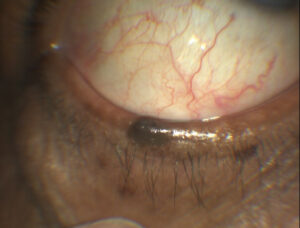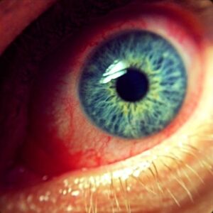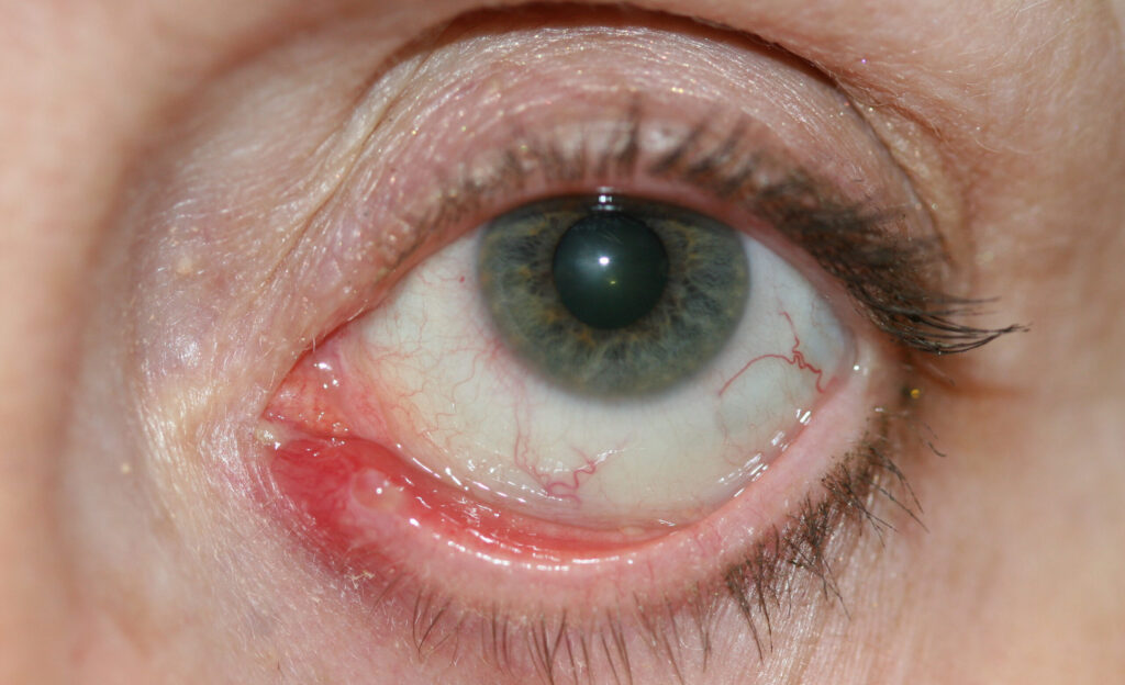
Canaliculitis is a chronic, often smoldering infection of the lacrimal canaliculus that is frequently misdiagnosed as ordinary chronic conjunctivitis, and the two conditions can look deceptively similar for months before the correct diagnosis is finally made, usually only after a course of standard conjunctivitis treatment has failed and someone specifically presses on the canalicular region and produces the characteristic discharge that gives the diagnosis away.
Why It’s So Often Missed
The chronic mucopurulent discharge and mild conjunctival injection produced by canaliculitis closely resemble ordinary bacterial or chronic conjunctivitis, and unless the examiner specifically looks at and palpates the canalicular region near the medial canthus — rather than focusing purely on the conjunctiva and lid margins more broadly — the actual source of the problem is easily overlooked.
A history of a chronic, unilateral, treatment-resistant “conjunctivitis” is itself a clue that should prompt specific examination of the punctum and canaliculus.
Because the two conditions are so often confused, patients frequently arrive having already tried multiple courses of topical antibiotic drops for presumed conjunctivitis, and this pattern of repeated, ineffective treatment is itself one of the more reliable signals pointing toward the correct diagnosis.
Microbiology
Actinomyces species (Actinomyces israelii), a filamentous, Gram-positive anaerobic bacterium, is the classic and most historically emphasized cause, notable for forming characteristic sulfur granules within the canaliculus that both help confirm the diagnosis microscopically and explain why the condition can be so persistent, since these concretions can shelter the organism from antibiotics that would otherwise be expected to clear the infection.
Other bacteria (staphylococcal and streptococcal species), as well as fungal and, less commonly, viral (herpes simplex) causes, have also been implicated, so a broader microbial spectrum should be kept in mind rather than assuming Actinomyces is responsible in every case.
This microbiologic variability is part of why culture results, when obtainable, genuinely change management rather than being a formality performed only to confirm an already-assumed diagnosis.
All-fit Slit-Lamp Adapter
Record and share exactly what you see at the slit lamp. One adapter fits any slit lamp or surgical microscope — and any smartphone.
From Choroida — the team behind this siteClinical Findings
- Chronic, often unilateral, mucopurulent discharge and mild conjunctival injection, easily mistaken for ordinary conjunctivitis
- Mild swelling and erythema localized specifically over the canalicular region, near the medial canthus — a subtle but genuinely useful localizing sign distinguishing this from more generalized conjunctivitis
- Expression of purulent material, sometimes containing visible small, yellowish “sulfur granules,” on gentle pressure over the affected canaliculus — a maneuver that, when performed and positive, is highly suggestive of the diagnosis
- A pouting, dilated, sometimes erythematous punctum on careful inspection, itself another subtle physical clue distinguishing this from simple conjunctivitis
- Failure to respond to standard topical antibiotics used for presumed ordinary conjunctivitis, which is often what eventually prompts reconsideration of the diagnosis
Differential Diagnosis
- Chronic bacterial or viral conjunctivitis — the most common misdiagnosis, distinguished by the absence of localized canalicular swelling and expressible discharge on canalicular pressure
- Dacryocystitis — swelling and infection centered over the lacrimal sac, below the medial canthal tendon, rather than the canaliculus itself, discussed in its own dedicated article on this site; the two structures are anatomically adjacent but distinct, and the location of maximal swelling and tenderness usually distinguishes them
- Punctal or canalicular foreign body or plug complication — a retained punctal plug, sometimes placed for dry eye management, can become a nidus for chronic infection resembling primary canaliculitis, and a history of prior plug placement should be specifically asked about
- Allergic conjunctivitis — itchy, typically bilateral, without the localized canalicular findings or expressible discharge characteristic of canaliculitis
Diagnostic Evaluation
The diagnosis is largely clinical, based on the characteristic history of chronic, treatment-resistant “conjunctivitis” combined with the exam finding of expressible discharge on canalicular pressure, a maneuver that should be part of the routine exam whenever this diagnosis is being considered.
Expressed material can be sent for Gram stain and culture, with a specific request for anaerobic culture given the possibility of Actinomyces, which requires particular culture conditions and enough incubation time that standard aerobic cultures alone can miss it, to confirm the causative organism and guide antimicrobial choice, a request the microbiology lab needs explicitly rather than assuming from a routine ocular specimen submission.
Management
Canaliculotomy — surgical opening of the canaliculus with mechanical removal of any concretions or granules — combined with irrigation is generally required for durable resolution, because the concretions characteristic of actinomycotic canaliculitis in particular can shelter the organism from antibiotics delivered by topical or even systemic routes alone.
Topical or systemic antibiotics are used adjunctively, tailored to culture results when available, but are frequently insufficient as monotherapy when concretions are present, which is why the surgical component of treatment is so central to actually curing the condition rather than just temporarily suppressing symptoms and allowing them to recur once treatment stops.
Warm compresses and gentle canalicular massage can help express material and provide some symptomatic relief while definitive treatment is arranged, and recurrence after adequate canaliculotomy and concretion removal is uncommon, making this generally a curable condition once correctly diagnosed and properly treated.
This favorable, curable prognosis is worth emphasizing to patients who may have spent months frustrated by a “conjunctivitis” that simply would not resolve with conventional treatment, since correctly identifying and treating canaliculitis typically resolves symptoms definitively rather than requiring the ongoing, repeated treatment they may have already experienced.


Document what you see
Two smartphone imaging tools built for everyday clinic use — one for the slit lamp, one for the fundus.
From Choroida — the team behind this siteReferences
- Freedman JR, Markert MS, Cohen AJ. Primary and secondary lacrimal canaliculitis: a review of literature. Survey of Ophthalmology.
- Vécsei VP, Huber-Spitzy V, Arocker-Mettinger E, Steinkogler FJ. Canaliculitis: difficulties in diagnosis, clinical features and treatment. Ophthalmologica.
- American Academy of Ophthalmology. Basic and Clinical Science Course, Section 7: Orbit, Eyelids, and Lacrimal System.
Test yourself
A few questions straight from this article.
-
Canaliculitis is a chronic infection centred on which structure?
Canaliculitis is a chronic, smouldering infection of the lacrimal canaliculus, which is why swelling and tenderness localise near the medial canthus rather than diffusely across the conjunctiva. -
Which organism is the classic and most historically emphasised cause of canaliculitis?
Actinomyces israelii, a filamentous Gram-positive anaerobic bacterium, is the classic cause and forms the characteristic sulfur granules seen within the canaliculus. -
Why does actinomycotic canaliculitis persist despite appropriate antibiotics?
The concretions formed inside the canaliculus protect the organism from antibiotics delivered topically or systemically, which is why drugs alone so often fail to cure the infection. -
What is expressed on gentle pressure over the canaliculus in canaliculitis?
Gentle pressure over the affected canaliculus expresses purulent material that sometimes contains visible small yellowish sulfur granules; a positive result is highly suggestive of the diagnosis. -
Which punctal appearance is a subtle clue to canaliculitis on careful inspection?
A pouting, dilated and sometimes erythematous punctum is another subtle physical clue that separates canaliculitis from simple conjunctivitis. -
What feature distinguishes dacryocystitis from canaliculitis on examination?
Dacryocystitis centres on the lacrimal sac below the medial canthal tendon, whereas canaliculitis localises to the canaliculus itself; the site of maximal swelling and tenderness usually separates them. -
Which prior intervention should be asked about as a cause of secondary canaliculitis?
A retained punctal plug, often placed for dry eye management, can become a nidus for chronic infection that mimics primary canaliculitis, so prior plug placement should be specifically asked about. -
What must be specifically requested when sending expressed canalicular material to the laboratory?
Anaerobic culture must be requested explicitly because Actinomyces needs particular culture conditions and sufficient incubation time, and standard aerobic cultures alone can miss it. -
What treatment is generally required for durable resolution of canaliculitis?
Surgical opening of the canaliculus with mechanical removal of concretions, combined with irrigation, is generally needed because antibiotics alone cannot reach organisms sheltered inside the concretions. -
What is the expected outcome after adequate canaliculotomy and concretion removal in canaliculitis?
Recurrence after adequate canaliculotomy with concretion removal is uncommon, making canaliculitis generally curable once it is correctly diagnosed and properly treated.

Writes and reviews the clinical articles on Educate. Every post is checked against peer-reviewed sources before it goes live.