
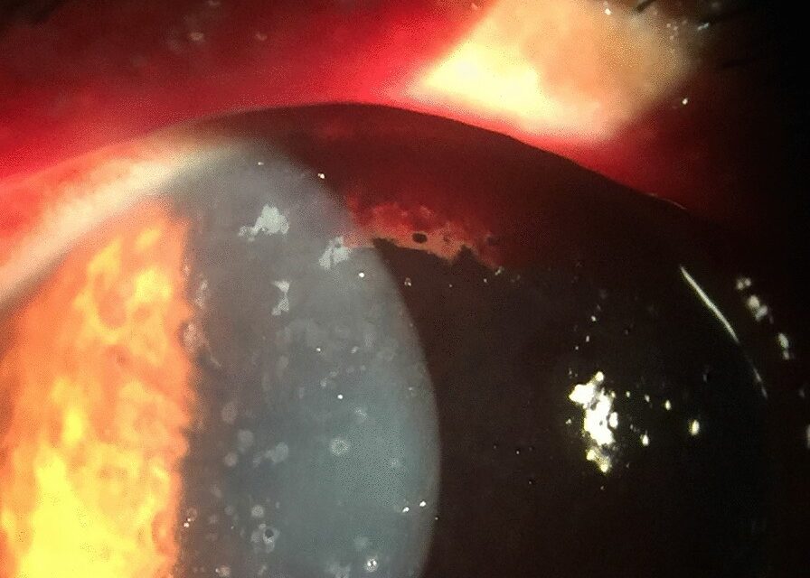
Microsporidial keratoconjunctivitis is an infection of the corneal and conjunctival epithelium by obligate intracellular microsporidian organisms, and it produces coarse punctate epithelial lesions that are frequently mistaken for adenoviral keratoconjunctivitis or dry eye.
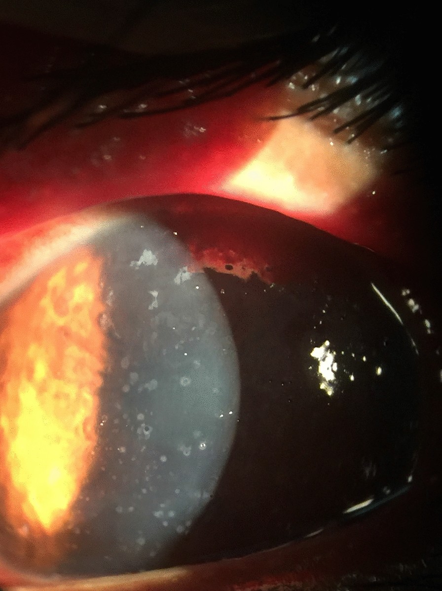
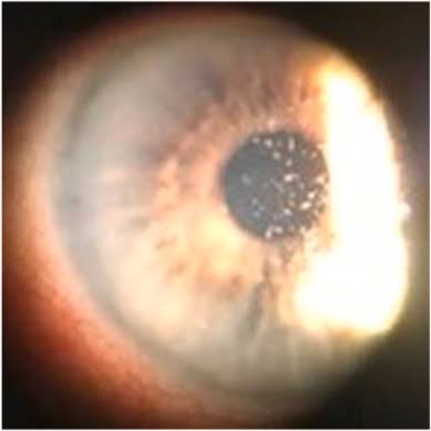
Two distinct clinical patterns exist, one in immunocompromised patients with disseminated disease and one in otherwise healthy contact lens wearers or after minor trauma, and telling them apart changes how aggressively systemic disease is pursued.
The Organism
Microsporidia are obligate intracellular fungi-related organisms, and several genera cause human ocular disease, most often Encephalitozoon and Vittaforma species.
Infection occurs through direct inoculation of the ocular surface, from contaminated water, soil, or contact lens solutions, or as part of disseminated infection in patients with advanced immunosuppression.
Two Clinical Patterns
Immunocompromised Patients
In patients with advanced HIV infection or other severe immunosuppression, microsporidial keratoconjunctivitis presents as a chronic, bilateral, diffuse punctate epitheliopathy.
- Coarse, gray-white, raised epithelial lesions scattered across the cornea
- Conjunctival injection and mild discharge
- Symptoms of foreign body sensation, redness, and photophobia rather than severe pain
- Disease is often part of disseminated microsporidiosis, with intestinal, respiratory, or renal involvement
Immunocompetent Patients
A separate, usually self-limited stromal keratitis pattern occurs in healthy individuals, often linked to trauma, soil or water exposure, or contact lens wear.
This form can produce deeper stromal infiltrates and is managed differently from the epithelial disease of immunocompromised patients.
All-fit Slit-Lamp Adapter
Record and share exactly what you see at the slit lamp. One adapter fits any slit lamp or surgical microscope — and any smartphone.
From Choroida — the team behind this siteDiagnosis
Microsporidial keratoconjunctivitis is easy to miss because the coarse punctate pattern resembles several more common conditions.
- Corneal scraping with special stains, including modified trichrome and Gram chromotrope, which highlight the small intracellular spores
- Confocal microscopy, which can show the characteristic oval intraepithelial organisms in vivo
- PCR testing where available, for species identification
- In immunocompromised patients, evaluation for disseminated disease, since ocular involvement may be the presenting sign of a systemic infection
A persistent, bilateral punctate keratoconjunctivitis in a patient with advanced HIV infection should prompt specific testing for microsporidia rather than a default diagnosis of viral keratoconjunctivitis.
Differential Diagnosis
- Adenoviral keratoconjunctivitis, which is usually more acute and self-limited
- Thygeson superficial punctate keratitis, which has a different clinical course and does not occur specifically in immunocompromised patients (see Thygeson superficial punctate keratitis)
- Dry eye disease
- Chlamydial or other atypical conjunctivitis
Management
Topical Therapy
Fumagillin eye drops, when available, are the most specific treatment and are effective against the epithelial disease seen in immunocompromised patients.
Topical antiseptics such as propamidine isethionate and polyhexamethylene biguanide have also been used, particularly for the stromal form in immunocompetent patients, borrowing from Acanthamoeba treatment protocols.
Systemic Therapy
Oral albendazole is used, particularly for patients with disseminated microsporidiosis, and it can help reduce the systemic organism burden even when the ocular response to topical therapy alone is incomplete.
Immune Reconstitution
In HIV-positive patients, restoring immune function with effective antiretroviral therapy is central to resolving the infection, since the ocular disease often persists as long as the underlying immunosuppression continues.
Prognosis
The epithelial disease of immunocompromised patients often persists until immune function improves, even with appropriate topical treatment, while the stromal disease of immunocompetent patients tends to resolve with topical therapy over weeks.
Recognizing the organism early avoids a long, frustrating course of misdirected treatment for presumed viral or allergic conjunctivitis.


Document what you see
Two smartphone imaging tools built for everyday clinic use — one for the slit lamp, one for the fundus.
From Choroida — the team behind this siteReferences
- Font RL, Samaha AN, Keener MJ, Chevez-Barrios P, Goosey JD. Corneal microsporidiosis: report of case, including electron microscopic observations. Ophthalmology. 2003;110:1418-1422.
- Joseph J, Sharma S, Murthy SI, et al. Microsporidial keratitis in India: 16S rRNA gene-based PCR assay for diagnosis and species identification of microsporidia in clinical samples. Invest Ophthalmol Vis Sci. 2006;47:4468-4473.
- Das S, Sharma S, Sahu SK, Rath S, Sahoo PK. New microbiological insights into microsporidial keratoconjunctivitis. Ophthalmology. 2012;119:39-45.
- Vemuganti GK, Garg P, Sharma S, et al. Is microsporidial keratitis an emerging cause of stromal keratitis? A case series study. BMC Ophthalmol. 2005;5:19.
