
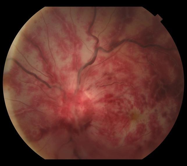
Ophthalmic artery occlusion is a more devastating variant of acute retinal arterial occlusion, distinguished from the more familiar central retinal artery occlusion by involving the ophthalmic artery itself — the parent vessel that supplies not only the central retinal artery but also the entire choroidal circulation via the posterior ciliary arteries.
Because both the inner retina (via the central retinal artery) and the outer retina and choroid (via the choroidal circulation) lose their blood supply simultaneously, the resulting vision loss is characteristically more profound and the visual prognosis considerably worse than in a typical, isolated central retinal artery occlusion.
Why Combined Involvement Matters
In isolated central retinal artery occlusion, the choroidal circulation remains intact, continuing to supply the outer retina and providing some degree of residual nutritional support and, in some cases, enough perfusion via collateral pathways (such as a cilioretinal artery, when present) to preserve a measure of central vision even amid severe overall visual loss.
When the ophthalmic artery itself is occluded, this choroidal reserve is lost along with the retinal circulation, removing the partial protective mechanism that sometimes limits the severity of vision loss in a simple CRAO and typically resulting in more complete, more severe, less recoverable vision loss.
Causes
- Embolic disease — from carotid atherosclerotic plaque or a cardiac source, the same general embolic mechanisms responsible for central retinal artery occlusion, but affecting a more proximal point in the arterial tree
- Giant cell arteritis — a critical cause to consider and urgently exclude in any older patient, given the immediate treatment implications and the risk of fellow-eye or other vascular territory involvement if untreated, discussed in more detail in this site’s coverage of arteritic anterior ischemic optic neuropathy
- Carotid artery dissection
- Orbital or periocular trauma, including, notably, iatrogenic cases following cosmetic facial filler injection, where inadvertent intravascular injection and retrograde embolization into the ophthalmic artery has become an increasingly recognized cause
- Hypercoagulable states and other less common causes of arterial occlusion
Fundus Explorer Pro
Photograph the retinal findings described here with the phone already in your pocket — 22 D optics and built-in illumination in one handheld unit.
From Choroida — the team behind this siteClinical Presentation
Patients present with sudden, severe, typically profound monocular vision loss, often more complete than the vision loss described by patients with an isolated central retinal artery occlusion.
Pain may accompany the event when the underlying cause involves an inflammatory process such as giant cell arteritis or an embolic event with associated ocular ischemia.
Given the severity and the differential that includes giant cell arteritis, associated systemic symptoms (headache, jaw claudication, scalp tenderness, malaise) should be specifically asked about, because their presence or absence meaningfully changes the urgency and nature of the workup.
Exam Findings
- Marked, often severe reduction in visual acuity, frequently worse than typically seen in isolated central retinal artery occlusion
- Diffuse retinal whitening/edema affecting essentially the entire posterior pole, reflecting combined retinal and choroidal ischemia rather than the retinal-only whitening of simple CRAO
- Absence of a cherry-red spot in many cases, or a less distinct one than classically described in isolated CRAO — since the cherry-red spot itself depends on relatively preserved choroidal perfusion showing through a thin fovea against surrounding opaque, ischemic retina, and this contrast is lost when the choroid is also ischemic
- A marked relative afferent pupillary defect, discussed in more detail in this site’s dedicated article on that sign
- Signs of associated anterior segment ischemia in more severe or prolonged cases, given the ophthalmic artery’s broader role in supplying anterior as well as posterior ocular structures
Differential Diagnosis
- Isolated central retinal artery occlusion — retinal whitening with a preserved, often prominent cherry-red spot, and generally somewhat less severe overall vision loss given intact choroidal perfusion
- Central retinal vein occlusion — a fundamentally different fundus appearance (widespread hemorrhage, disc edema, venous dilation) rather than the retinal whitening of arterial occlusion, and a different, though still often significant, degree and pattern of vision loss
- Vitreous hemorrhage or other media opacity — reduced vision without the characteristic fundus findings of arterial occlusion, generally distinguishable once adequate fundus visualization is obtained
Diagnostic Evaluation
Urgent evaluation for giant cell arteritis — inflammatory markers, temporal artery biopsy where clinical suspicion is significant — is essential in any older patient given the treatment urgency and the risk to the fellow eye if this diagnosis is missed and left untreated.
Carotid imaging and a cardiac embolic workup, following the same general principles used for any acute arterial retinal occlusion, are appropriate once giant cell arteritis has been reasonably addressed, given the shared embolic risk factors between ophthalmic and central retinal artery occlusion.
Management
As with central retinal artery occlusion generally, treatment options for established ophthalmic artery occlusion are limited and the evidence supporting any specific intervention (ocular massage, anterior chamber paracentesis, hyperbaric oxygen) is weak, reflecting the same fundamental challenge that affects management of acute retinal arterial occlusion overall.
Treatment is far more effective, in principle, before irreversible ischemic damage occurs rather than after, and if giant cell arteritis is suspected, immediate high-dose systemic corticosteroid therapy should not be delayed for biopsy confirmation, given the urgency of protecting the fellow eye from the same, often devastating, process.
Given the severity of vision loss and generally poor visual prognosis once ophthalmic artery occlusion has occurred, the greater practical impact often lies in the broader workup: identifying and addressing the underlying cause (giant cell arteritis, carotid disease, cardioembolic source) to prevent further vascular events, including stroke, which shares many of the same underlying risk factors and represents a serious, sometimes more immediately life-threatening, associated risk.


Document what you see
Two smartphone imaging tools built for everyday clinic use — one for the slit lamp, one for the fundus.
From Choroida — the team behind this siteReferences
- Hayreh SS. Ocular vascular occlusive disorders: natural history of visual outcome. Progress in Retinal and Eye Research.
- Beatty S, Au Eong KG. Acute occlusion of the retinal arteries: current concepts and recent advances in diagnosis and management. Journal of Accident & Emergency Medicine.
- American Academy of Ophthalmology. Basic and Clinical Science Course, Section 12: Retina and Vitreous.
