
Older patients sometimes show up with a small gray or blue-gray patch on the white of the eye, typically right in front of the insertion of the medial or lateral rectus muscle.
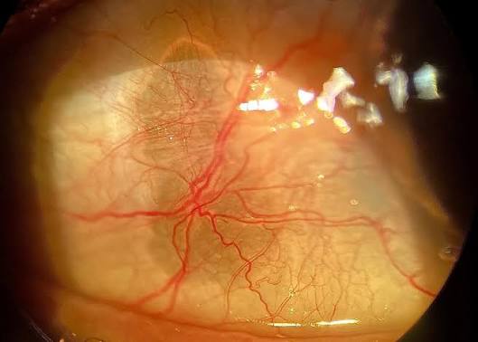
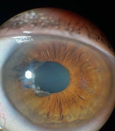
It is flat, smooth, and well-defined, and it often occurs on both sides.
This is a senile scleral plaque, and its main clinical interest is that it is benign.
What it is
The plaque is a patch of hyalinized, calcified sclera in the area where the rectus tendon inserts.
The tendon places repeated mechanical stress on the sclera over decades, and degeneration, with calcium and sometimes hyaline deposition, occurs.
The sclera in that zone becomes translucent, and the darker uvea beneath shows through as a gray or bluish tint, which is why it can be mistaken for thinning or a pigmented lesion.
It is a standard topic in ocular histopathology.
Appearance
- A well-demarcated, oval or crescent-shaped patch just anterior to the muscle insertion
- Gray, blue-gray, or slate color that can look like pigmentation
- Smooth surface without elevation, and normal conjunctiva and episclera over it
- Most often horizontal and bilateral, nasal more than temporal
- Found in people over 60, and more common with increasing age
It does not bulge, does not change in size, and is not painful.
All-fit Slit-Lamp Adapter
Record and share exactly what you see at the slit lamp. One adapter fits any slit lamp or surgical microscope — and any smartphone.
From Choroida — the team behind this siteDifferential diagnosis
- Scleral thinning from scleritis, which is painful, inflamed, and involves a darker, bluish area with vessels that are often engorged (see necrotizing scleritis and scleromalacia perforans)
- Scleral staphyloma, where the uvea bulges forward
- Epibulbar melanocytic lesions that are brown or black and sometimes elevated
- Scleral calcium deposition in hypercalcemia or hyperphosphatemia, which can be inflamed
- Scleral changes in connective tissue disorders such as osteogenesis imperfecta, in which the whole sclera appears blue
The age of the patient, the position, and the lack of inflammation point to a plaque.
Do you need any tests?
Usually not.
In a patient with an atypical appearance, or if there is pain or redness, imaging with anterior segment OCT can confirm that scleral thickness is preserved.
Systemic evaluation for calcium disorders is warranted only if there are other clues.
What to tell the patient
It is a normal ageing change, it is not a growth, and it does not cause trouble.
The patient should be told that it will not change significantly, and that they should come back if the area becomes red or painful, or starts to bulge.
Management
No treatment is needed.
Surgery, even for cosmetic reasons, is not indicated, because it carries a risk of perforation and offers little benefit.
Where exactly it occurs
Plaques lie in the interpalpebral zone, just anterior to the insertion of the horizontal rectus tendons, often described as being 2 to 4 mm wide and 4 to 7 mm long, with a convex margin facing the cornea. They are often seen on the nasal side, and may be symmetrical. The sclera in the plaque is not thinned in the sense of a staphyloma, but it has become translucent from hyalinization and calcium deposition, so the darker tissue beneath shows through.
How to separate it from true scleral thinning
In scleritis, the area is red, tender, and sometimes bulging, with engorged vessels in the episclera and sclera. In a plaque, the conjunctiva and episclera are normal, the surface is flat, the patient has no pain, and the color is a stable slate gray. If there is doubt, anterior segment OCT shows that the scleral thickness is preserved, and the patient can be reviewed in a few weeks.
Reassurance and records
Explain that the plaque is a sign of ageing of the sclera, that it does not cause trouble, and that it does not mean that the eye wall is weak. Document with a photograph and a note that the appearance is typical, so that the finding is not mistaken for disease at a later visit. Avoid any procedure that cuts or cauterizes the area.
Associations
Scleral plaques are more common with advancing age and are slightly more frequent in women. They are not linked with any systemic disease. Similar changes in younger patients should prompt a look for other causes, such as connective tissue disorders or calcium and phosphate abnormalities, but in older patients with a typical appearance, no tests are needed.
Surgical relevance
The plaque area can be thinner than the surrounding sclera, so surgeons should be careful with sutures and cautery near the horizontal rectus insertions, for example in strabismus or retinal detachment surgery.
Prognosis
Excellent.
The plaque is purely cosmetic, and may be noted in the chart for future reference.


Document what you see
Two smartphone imaging tools built for everyday clinic use — one for the slit lamp, one for the fundus.
From Choroida — the team behind this siteReferences
- Bron AJ, Tripathi RC, Tripathi BJ. Wolff’s Anatomy of the Eye and Orbit. 8th ed. London: Chapman & Hall; 1997.
- Watson PG, Young RD. Scleral structure, organisation and disease. A review. Exp Eye Res. 2004;78:609-623.
- Kanski JJ, Bowling B. Clinical Ophthalmology: A Systematic Approach. 7th ed. London: Elsevier; 2011.
- Fine BS, Yanoff M. Ocular Histology: A Text and Atlas. 2nd ed. New York: Harper & Row; 1979.
