
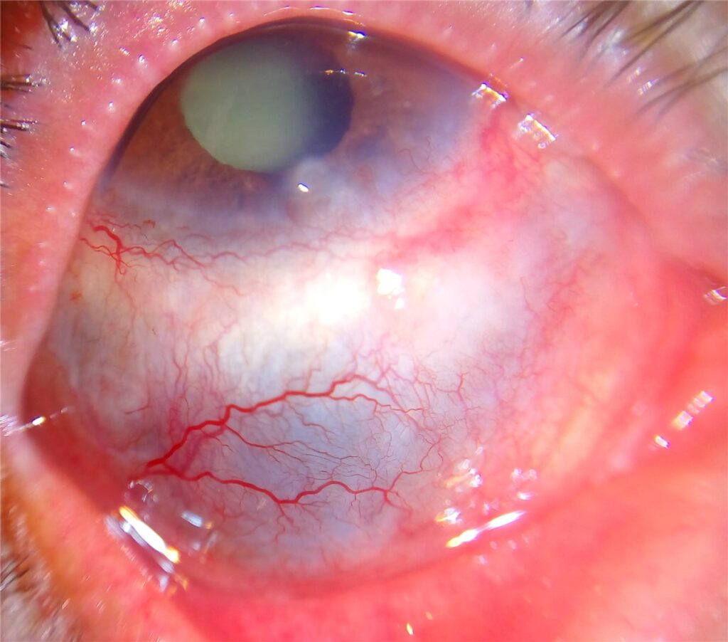
Granulomatosis with polyangiitis, formerly called Wegener’s granulomatosis, is a systemic small-vessel vasculitis affecting the respiratory tract and kidneys, and the eye is involved often enough that ophthalmic disease is sometimes the presenting feature that leads to the systemic diagnosis, not simply a complication that follows it.
Necrotizing scleritis is the finding most closely associated with this disease, and it carries real urgency: scleritis of this severity, left inadequately treated, can perforate the globe, and the systemic vasculitis behind it can be fatal without appropriate immunosuppression.
Systemic Disease Overview
Granulomatosis with polyangiitis is characterised by necrotizing granulomatous inflammation and vasculitis affecting small to medium vessels, classically involving the upper respiratory tract, lungs, and kidneys.
ANCA-associated vasculitis, most often with a cytoplasmic (c-ANCA) pattern targeting proteinase 3, is present in the majority of patients with active systemic disease, and its identification is a central part of both diagnosis and disease monitoring.
Ocular Manifestations
Necrotizing Scleritis
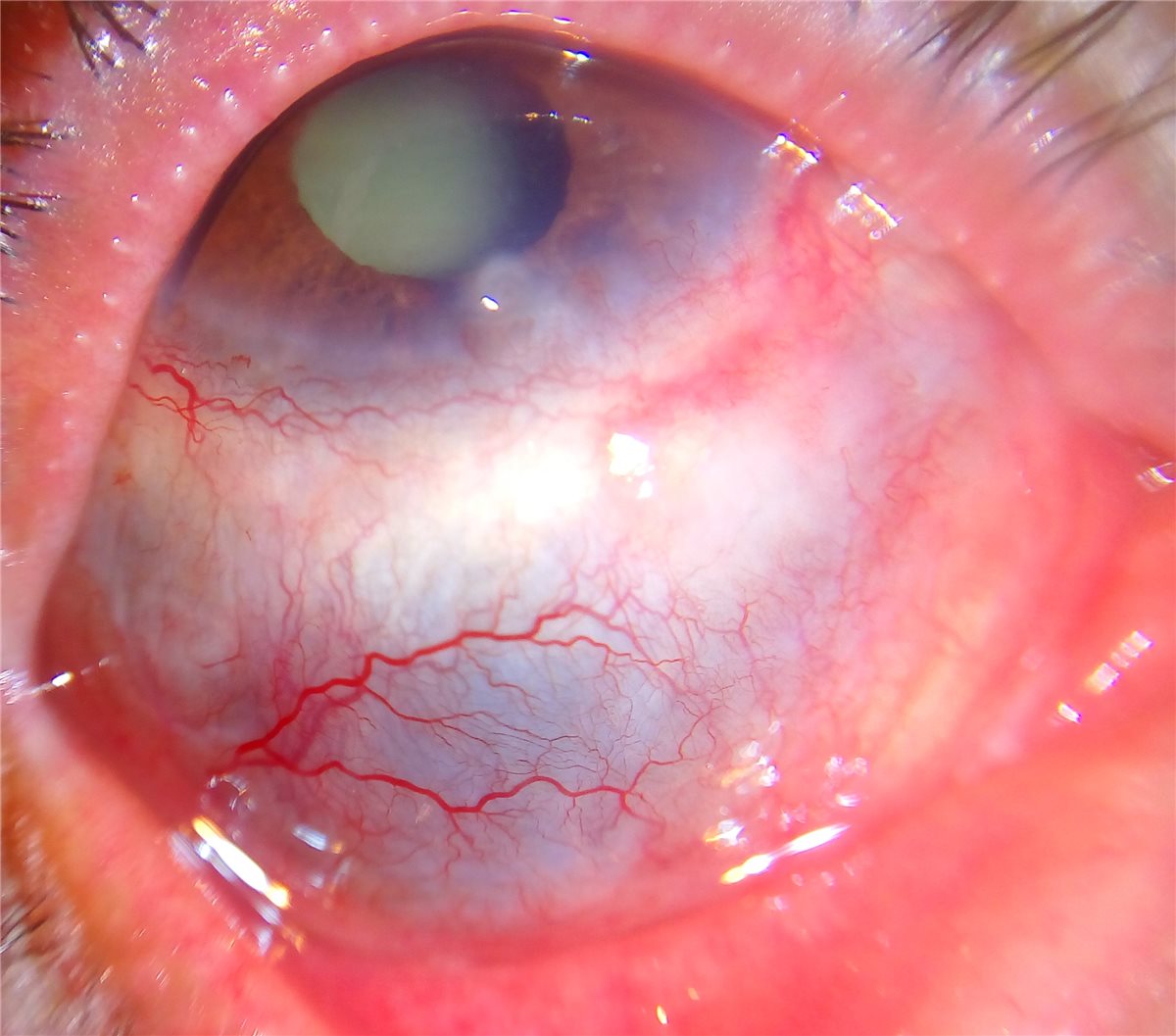
Severe, often bilateral scleritis with a tendency toward scleral necrosis and thinning is one of the most characteristic and sight-threatening ocular manifestations, and necrotizing scleritis in any patient should prompt screening for an underlying systemic vasculitis, of which this is among the most important causes.
Peripheral Ulcerative Keratitis
Progressive, crescentic thinning and ulceration of the peripheral cornea can occur alone or alongside scleritis, and when severe is associated with significant risk of perforation, making it one of the more urgent ocular presentations in this disease.
Orbital Pseudotumour
Granulomatous orbital inflammation can cause proptosis, restricted eye movement, and compressive optic neuropathy, and orbital involvement in this disease is often more destructive and less steroid-responsive than idiopathic orbital inflammation, frequently requiring more aggressive systemic immunosuppression.
Other Findings
Nasolacrimal duct obstruction from involvement of the nasal mucosa, uveitis, and retinal vasculitis are all described, and any combination of ocular inflammation alongside sinonasal or pulmonary symptoms should raise suspicion for this diagnosis.
All-fit Slit-Lamp Adapter
Record and share exactly what you see at the slit lamp. One adapter fits any slit lamp or surgical microscope — and any smartphone.
From Choroida — the team behind this siteSystemic Findings
- Upper respiratory tract involvement, including chronic sinusitis, nasal crusting, epistaxis, and, in severe cases, saddle nose deformity from cartilage destruction
- Pulmonary involvement, including nodules, cavitary lesions, and pulmonary haemorrhage
- Renal involvement, classically a rapidly progressive glomerulonephritis, which is a major driver of morbidity and mortality if untreated
- Constitutional symptoms, including fever, weight loss, and malaise, common at presentation
Diagnostic Evaluation
ANCA testing, specifically c-ANCA with anti-proteinase 3 specificity, supports the diagnosis in the appropriate clinical context, though a negative result does not fully exclude the disease, particularly in limited or localised presentations.
Tissue biopsy, from an affected site such as the sinuses, lung, or kidney, showing necrotizing granulomatous vasculitis provides the most definitive confirmation, though a compatible clinical picture with positive ANCA serology is often sufficient to begin treatment without biopsy when the presentation is classic.
Renal function testing and urinalysis for proteinuria or haematuria are essential given the frequency and seriousness of renal involvement, and CT imaging of the chest and sinuses helps characterise the extent of systemic disease.
Differential Diagnosis
- Rheumatoid arthritis-associated scleritis, generally occurring in a patient with an established history of joint disease rather than as a first presentation of systemic vasculitis
- Other ANCA-associated vasculitides, including microscopic polyangiitis and eosinophilic granulomatosis with polyangiitis, distinguished by their own characteristic organ involvement patterns and, in the latter, prominent asthma and eosinophilia
- Relapsing polychondritis, which can cause scleritis alongside cartilage inflammation elsewhere, distinguished by involvement of the ear and nasal cartilage rather than the sinonasal and renal pattern of GPA
- Infectious scleritis, generally with a relevant history of trauma or surgery and a different, more localised clinical course
Management
Systemic Immunosuppression
Treatment of the underlying systemic vasculitis, typically with corticosteroids combined with cyclophosphamide or rituximab for remission induction, followed by maintenance immunosuppression, is essential and is managed jointly with rheumatology, since the ocular disease will not resolve without control of the underlying systemic process.
Local Ocular Treatment
Topical treatment alone is inadequate for necrotizing scleritis or peripheral ulcerative keratitis in this disease; systemic immunosuppression is required, with topical lubrication and cycloplegia used as adjuncts for comfort rather than as primary therapy.
Surgical Considerations
Impending or actual scleral or corneal perforation may require surgical reinforcement with a patch graft, and any surgery should ideally be undertaken once systemic inflammation is at least partially controlled, since operating on actively inflamed tissue in this disease carries a high risk of poor wound healing and further breakdown.
Prognosis
Untreated systemic granulomatosis with polyangiitis has a poor prognosis, historically associated with high mortality within the first year, primarily from renal and pulmonary involvement, though modern immunosuppressive regimens have transformed this outlook substantially.
Ocular prognosis depends heavily on how promptly systemic immunosuppression is started relative to the severity of scleral or corneal involvement, since necrotizing scleritis and peripheral ulcerative keratitis can each progress to perforation and vision loss if treatment is delayed.
With appropriate systemic treatment, many patients achieve remission of both ocular and systemic disease, though relapse is common and lifelong monitoring, coordinated between ophthalmology and rheumatology, remains necessary even after apparent disease control.


Document what you see
Two smartphone imaging tools built for everyday clinic use — one for the slit lamp, one for the fundus.
From Choroida — the team behind this siteReferences
- Pakrou N, Selva D, Leibovitch I. Wegener’s granulomatosis: ophthalmic manifestations and management. Seminars in Arthritis and Rheumatism. 2006.
- Rothschild PR, Pagnoux C, Seror R, et al. Ophthalmologic manifestations of systemic necrotizing vasculitides at diagnosis. Journal of Rheumatology. 2013.
- Kubaisi B, Abu Samra K, Foster CS. Granulomatosis with polyangiitis (Wegener’s disease): an updated review of ocular disease manifestations. Intractable & Rare Diseases Research. 2016.
- Granulomatosis with Polyangiitis. EyeWiki, American Academy of Ophthalmology.
- Granulomatosis with Polyangiitis. StatPearls, NCBI Bookshelf.
Test yourself
A few questions straight from this article.
-
Which organ systems are classically involved in granulomatosis with polyangiitis?
The disease is a necrotizing granulomatous small- to medium-vessel vasculitis that classically targets the upper airway, lungs and kidneys. -
Which serological pattern is characteristic of granulomatosis with polyangiitis?
A cytoplasmic ANCA pattern with anti-proteinase 3 specificity is present in the majority of patients with active systemic disease and is central to diagnosis and monitoring. -
What should the finding of necrotizing scleritis prompt in any patient?
Necrotizing scleritis is strongly associated with systemic vasculitis, of which granulomatosis with polyangiitis is among the most important causes, so it should trigger a systemic search. -
Which corneal complication of granulomatosis with polyangiitis carries a high risk of perforation?
Progressive crescentic thinning and ulceration of the peripheral cornea can occur with or without scleritis and is one of the more urgent ocular presentations of the disease. -
How does orbital involvement in granulomatosis with polyangiitis differ from idiopathic orbital inflammation?
Granulomatous orbital inflammation in this disease tends to be more destructive and less steroid-responsive, frequently requiring more aggressive systemic immunosuppression. -
What provides the most definitive confirmation of granulomatosis with polyangiitis?
Tissue from an affected site such as sinus, lung or kidney showing necrotizing granulomatous vasculitis is the most definitive confirmation, though a classic picture with positive ANCA often suffices to start treatment. -
Which systemic testing is essential in suspected granulomatosis with polyangiitis?
Renal function and urinalysis for proteinuria or haematuria are essential given the frequency and seriousness of the rapidly progressive glomerulonephritis this disease causes. -
What distinguishes relapsing polychondritis from granulomatosis with polyangiitis as a cause of scleritis?
Relapsing polychondritis causes scleritis alongside cartilage inflammation of the ear and nose, rather than the sinonasal and renal pattern that characterises granulomatosis with polyangiitis. -
What is required to control necrotizing scleritis in granulomatosis with polyangiitis?
Topical treatment alone is inadequate; corticosteroids with cyclophosphamide or rituximab are used for remission induction, since the eye will not settle until the systemic vasculitis is controlled. -
What is the outlook for untreated systemic granulomatosis with polyangiitis?
Untreated disease historically carried high first-year mortality driven by renal and pulmonary involvement, an outlook transformed by modern immunosuppressive regimens.

Writes and reviews the clinical articles on Educate. Every post is checked against peer-reviewed sources before it goes live.


