
Case Study
A 65-year-old male with a history of poorly controlled hypertension and diabetes mellitus presented to an ophthalmology clinic with a gradual loss of vision in his right eye over several months.
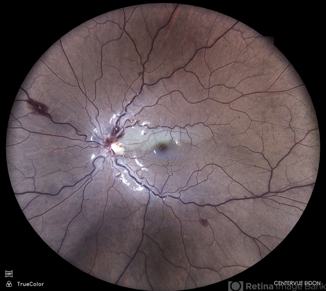
The patient described a decrease in visual acuity and noted occasional episodes of blurred vision. He denied any eye pain or photophobia.
His medical history was significant for peripheral vascular disease and previous transient ischemic attacks (TIAs).
Examination Results:
- Visual Acuity: 20/60 in the right eye and 20/30 in the left eye.
- Fundus Examination: Dilated and tortuous retinal veins with intraretinal hemorrhages and microaneurysms, particularly in the mid-peripheral retina of the right eye.
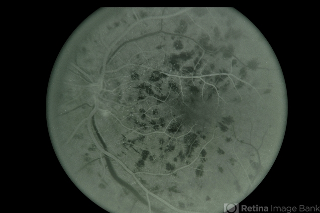
- Fluorescein Angiography: Delayed venous filling and extensive capillary nonperfusion in the right eye.
- Optical Coherence Tomography (OCT): Retinal thickening with cystoid macular edema in the right eye.
- Intraocular Pressure (IOP): 18 mmHg in both eyes.
A diagnosis of Venous Stasis Retinopathy was made.
Venous stasis retinopathy Disease entity
Venous Stasis Retinopathy is a retinal vascular disorder associated with chronic venous insufficiency, leading to impaired retinal blood flow and subsequent retinal ischemia.
The condition is characterized by the presence of dilated and tortuous retinal veins, intraretinal hemorrhages, and areas of capillary nonperfusion.
Venous stasis retinopathy is often associated with systemic vascular diseases such as hypertension, diabetes mellitus, and atherosclerosis.
It can be considered a precursor to central retinal vein occlusion (CRVO) but with a more insidious onset and a gradual progression.
This condition is part of a spectrum of diseases resulting from chronic venous stasis in the retina. Unlike CRVO, where venous occlusion is sudden and complete, venous stasis retinopathy develops more slowly, with partial obstruction of venous outflow over time.
This leads to chronic low-grade ischemia, which progressively damages the retinal tissue. Understanding the underlying mechanisms, clinical features, and appropriate management strategies is crucial for preventing severe visual loss in affected patients.
Venous stasis retinopathy is often underdiagnosed due to its subtle presentation and gradual progression. However, early recognition and management can significantly impact the patient’s visual prognosis.
Fundus Explorer Pro
Photograph the retinal findings described here with the phone already in your pocket — 22 D optics and built-in illumination in one handheld unit.
From Choroida — the team behind this sitePathophysiology
The pathophysiology of venous stasis retinopathy is primarily related to chronic venous insufficiency, which results in impaired venous outflow from the retina. This venous congestion leads to increased intraluminal pressure within the retinal veins, causing them to dilate and become tortuous.
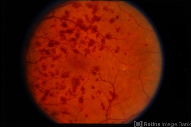
The increased pressure and reduced blood flow also contribute to endothelial damage and breakdown of the blood-retinal barrier, resulting in leakage of blood and plasma into the retinal tissue.
Over time, this chronic low-grade ischemia leads to retinal hypoxia, stimulating the production of vascular endothelial growth factor (VEGF) and other pro-angiogenic factors.
The retinal ischemia in venous stasis retinopathy often results in capillary nonperfusion, microaneurysm formation, and intraretinal hemorrhages.
If left untreated, the condition may progress to more severe forms of retinal vascular disease, including neovascularization and eventual retinal detachment.
The slow and insidious nature of venous stasis retinopathy often allows it to go unnoticed until significant retinal damage has occurred.
Venous stasis retinopathy Epidemiology
Venous stasis retinopathy is more commonly seen in older adults, particularly those with underlying systemic vascular diseases such as hypertension, diabetes, and atherosclerosis.
The condition is more prevalent in individuals with a history of peripheral vascular disease or cerebrovascular events, such as TIAs or strokes.
It tends to occur more frequently in individuals over the age of 50 and is slightly more common in males than females.
The exact prevalence of venous stasis retinopathy is difficult to determine, as the condition is often underdiagnosed or mistaken for other retinal vascular disorders.
However, it is believed to be a relatively common cause of gradual vision loss in older adults, particularly those with poorly controlled systemic vascular risk factors.
Clinical Features
Patients with venous stasis retinopathy typically present with a gradual decrease in visual acuity in one or both eyes.
The condition is usually painless and may go unnoticed until significant vision loss has occurred. Common clinical features include:
- Blurred Vision: Gradual onset of blurry vision, often worse in one eye.
- Visual Field Defects: Peripheral vision loss or scotomas may be present due to areas of capillary nonperfusion.
- Floaters: Occasional floaters may be reported due to intraretinal hemorrhages.
- Photopsia: Some patients may experience flashes of light due to retinal ischemia.
Venous stasis retinopathy Examination Findings
- Fundus Examination: Reveals dilated and tortuous retinal veins, intraretinal hemorrhages, microaneurysms, and areas of capillary nonperfusion.
- Fluorescein Angiography: Demonstrates delayed venous filling, capillary dropout, and areas of nonperfusion.
- Optical Coherence Tomography (OCT): Shows retinal thickening, cystoid macular edema, and sometimes subretinal fluid.
- Intraocular Pressure (IOP): Generally normal, but regular monitoring is essential to rule out secondary glaucoma.
Venous stasis retinopathy Differential Diagnosis
The differential diagnosis of venous stasis retinopathy includes several other retinal vascular disorders that present with similar clinical features:
- Central Retinal Vein Occlusion (CRVO): Sudden onset of vision loss with more extensive hemorrhages and optic disc swelling.
- Diabetic Retinopathy: Characterized by microaneurysms, hemorrhages, and neovascularization, typically associated with a history of diabetes.
- Hypertensive Retinopathy: Retinal changes associated with chronic hypertension, including arteriolar narrowing, hemorrhages, and cotton-wool spots.
- Branch Retinal Vein Occlusion (BRVO): Localized retinal vein occlusion affecting a sector of the retina, often with quadrant-specific findings.
Diagnosis
Diagnosis of venous stasis retinopathy is based on clinical examination and imaging studies:
- Fundus Examination: Key for identifying the characteristic retinal changes.
- Fluorescein Angiography: Critical for assessing the extent of capillary nonperfusion and venous filling defects.
- Optical Coherence Tomography (OCT): Provides detailed imaging of retinal edema and structural changes.
- Systemic Workup: Important for identifying and managing underlying systemic vascular risk factors.
Venous stasis retinopathy Management
Management of venous stasis retinopathy focuses on controlling the underlying systemic conditions and preventing further retinal damage:
- Control of Systemic Risk Factors: Tight control of blood pressure, blood sugar, and lipid levels is essential to reduce retinal ischemia.
- Anti-VEGF Therapy: Intravitreal injections of anti-VEGF agents may be used to reduce retinal edema and prevent neovascularization.
- Corticosteroids: Intravitreal corticosteroids may be considered in cases of persistent macular edema.
- Laser Photocoagulation: This may be used in cases of extensive capillary nonperfusion to reduce the risk of neovascularization.
- Regular Monitoring: Ongoing follow-up is essential to monitor disease progression and response to treatment.
Prognosis
The prognosis for venous stasis retinopathy varies depending on the severity of retinal ischemia and the effectiveness of treatment.
Early diagnosis and management of systemic risk factors can help slow the progression of the disease and preserve vision.
However, if left untreated, the condition may progress to more severe retinal complications, including neovascularization and retinal detachment, leading to significant vision loss.
Prevention
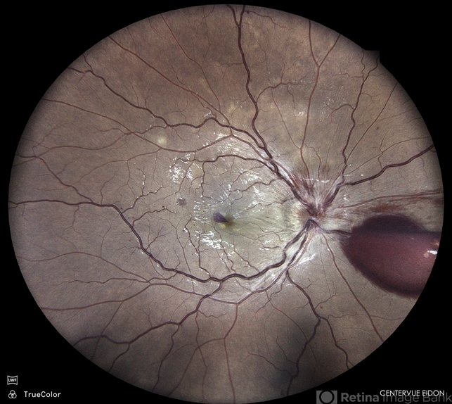
Prevention of venous stasis retinopathy involves the early identification and management of systemic vascular risk factors.
Public health efforts should focus on raising awareness about the importance of controlling hypertension, diabetes, and other vascular conditions to prevent retinal complications.
Regular eye examinations are crucial for early detection and management of venous stasis retinopathy, particularly in individuals with known systemic risk factors.
Conclusion
Venous stasis retinopathy is a significant retinal vascular disorder that can lead to gradual vision loss if not properly managed.
Understanding its pathophysiology, clinical features, and management strategies is essential for preventing severe visual impairment in affected patients.
Early intervention, coupled with effective management of systemic conditions, can improve outcomes and preserve vision.


Document what you see
Two smartphone imaging tools built for everyday clinic use — one for the slit lamp, one for the fundus.
From Choroida — the team behind this siteReferences
- Hayreh, S. S. (1994). Retinal vein occlusion. Indian Journal of Ophthalmology, 42(3), 109-132.
- McIntosh, R. L., Rogers, S. L., Lim, L., Cheung, N., Wang, J. J., Mitchell, P., & Wong, T. Y. (2010). Natural history of central retinal vein occlusion: an evidence-based systematic review. Ophthalmology, 117(6), 1113-1123.
- Rehak, M., & Wiedemann, P. (2010). Retinal vein thrombosis: pathogenesis and management. Journal of Thrombosis and Haemostasis, 8(9), 1886-1894.
- Scott, I. U., Flynn Jr, H. W., Smiddy, W. E., Murray, T. G., & Feuer, W. J. (1997).
Test yourself
A few questions straight from this article.
-
Venous stasis retinopathy is a retinal vascular disorder associated with which underlying process?
Venous stasis retinopathy arises from chronic venous insufficiency, which impairs retinal blood flow and causes retinal ischaemia with dilated tortuous veins, haemorrhages and capillary nonperfusion. -
How does venous stasis retinopathy relate to central retinal vein occlusion?
Venous stasis retinopathy can be considered a precursor to CRVO, but with a gradual, insidious course rather than the sudden complete occlusion seen in CRVO. -
What makes the retinal veins dilated and tortuous in venous stasis retinopathy?
Impaired venous outflow causes venous congestion and raises intraluminal pressure in the retinal veins, which dilate and become tortuous as a result. -
Untreated retinal hypoxia and VEGF release in venous stasis retinopathy risks which progression?
Chronic low-grade ischaemia drives VEGF production; if left untreated the disease may progress to neovascularisation and eventual retinal detachment with significant vision loss. -
What does fluorescein angiography characteristically show in venous stasis retinopathy?
Angiography in venous stasis retinopathy demonstrates delayed venous filling together with capillary dropout and areas of nonperfusion, which define the extent of ischaemia. -
Which optical coherence tomography finding is typical of venous stasis retinopathy?
OCT in venous stasis retinopathy shows retinal thickening and cystoid macular oedema, and sometimes subretinal fluid. -
Which presentation favours central retinal vein occlusion over venous stasis retinopathy?
CRVO presents with sudden vision loss, more extensive haemorrhages and optic disc swelling, whereas venous stasis retinopathy progresses slowly. Quadrantic haemorrhage suggests BRVO and arteriolar narrowing suggests hypertensive retinopathy. -
Intraocular pressure is usually normal in venous stasis retinopathy, so why is it monitored regularly?
Intraocular pressure is generally normal in venous stasis retinopathy, but regular measurement is essential to rule out secondary glaucoma. -
What forms the cornerstone of management in venous stasis retinopathy?
Management centres on controlling the underlying systemic vascular disease: tight control of blood pressure, blood sugar and lipid levels reduces retinal ischaemia and further damage. -
What is the stated indication for laser photocoagulation in venous stasis retinopathy?
Laser photocoagulation is reserved for eyes with extensive capillary nonperfusion, where it lowers the risk of subsequent neovascularisation.
